


Article
Comparison of Dermatoscopoi to Help You Choose the Right One
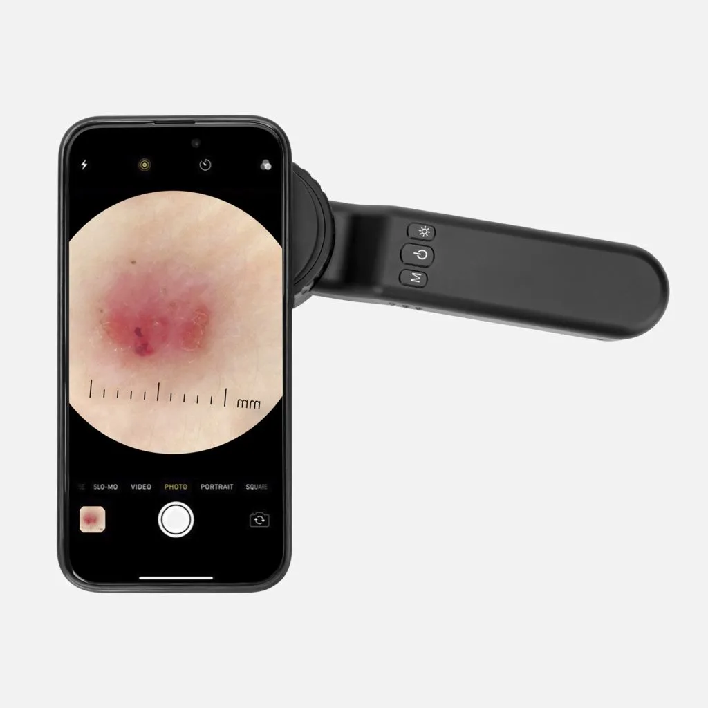
What is Dermoscopy (dermatoscopio)? Dermoscopy(dermatoscopio), is a valuable helpful technique usually used by skin doctors to inspect a variety of skin situations.Dermoscopy is also known as dermatoscopy, incident light microscopy, skin surface microscopy and epiluminescence microscopy. Dermoscopy commonly combines a high quality magnifying glass and a powerful lighting system. This greatly enhances the visual field of…
What is Dermoscopy (dermatoscopio)?
Dermoscopy(dermatoscopio), is a valuable helpful technique usually used by skin doctors to inspect a variety of skin situations.Dermoscopy is also known as dermatoscopy, incident light microscopy, skin surface microscopy and epiluminescence microscopy. Dermoscopy commonly combines a high quality magnifying glass and a powerful lighting system. This greatly enhances the visual field of subsurface skin structures and patterns that typically cannot be saw by the naked eye. The whole process for dermoscope inspection is non-invasive and painless. Dermoscopy is really a dependable and useful tool for skin examination.
Comparison of dermatoscopio:types
There are kinds of dermatoscopio, but most of them devided into two types, polarized dermoscopy and nonpolarized dermoscopy. In addition, there are new type named amber light dermoscopy springing up recent years. With the revolution of dermatoscope, a dermoscope typically with both nonpolarized light and polarized light. Some of them even with the amber light mode.
Comparison of dermatoscopio:basic principle
Nonpolarized dermoscope:
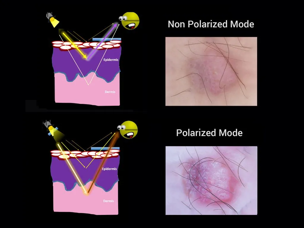
The oscillations of light waves of nonpolarized (unpolarized) light are in more than a single direction, like sunlight or light from lamps and torches. The waves from these objects oscillate in random directions.
Polarized dermoscope :
In polarized dermoscope, two polarized filters achieve orthogonally cross at 90 degrees. Vibration of polarized light is in a single plane. Polarized dermoscope can examine deeper layer of skin by eliminate reflection and glare on the ski surface.
Amber light dermoscope:
Amber light dermoscope is a specialized device illuminating the skin with amber-colored light.It utilizes a special wave length to highlight the visualization of certain features of the skin which are less visible by white light. It enhances not only the textures of skin lesions but also the contrast of surface features.
Comparison of dermatoscopio:clinical uses
Nonpolarized dermoscope:
Nonpolarized dermoscope is usually used for detecting structures of the superficial skin layer and inspecting the superficial epidermis down to the dermo-epidermal junction. It is better to assess surface characteristics and pigmented lesions.
Polarized Dermoscopy :
Polarized dermoscopy increases the visual of dermatologists to closely detect deeper skin layers, like dermo epidermal junction, superficial dermis, ect. It includes polarized dermoscopy contact and polarized dermoscopy non-contact.
Amber light Dermoscopy:
Under amber light dermoscope, by penetrating the certain depth of skin, it allows to detect the condition of structure below the top layer of epidermis. Amber light dermoscopy is better for diagnosing skin situations, such as vascular structures, pigmented areas,etc.
Comparison of dermatoscopio:ways of examination
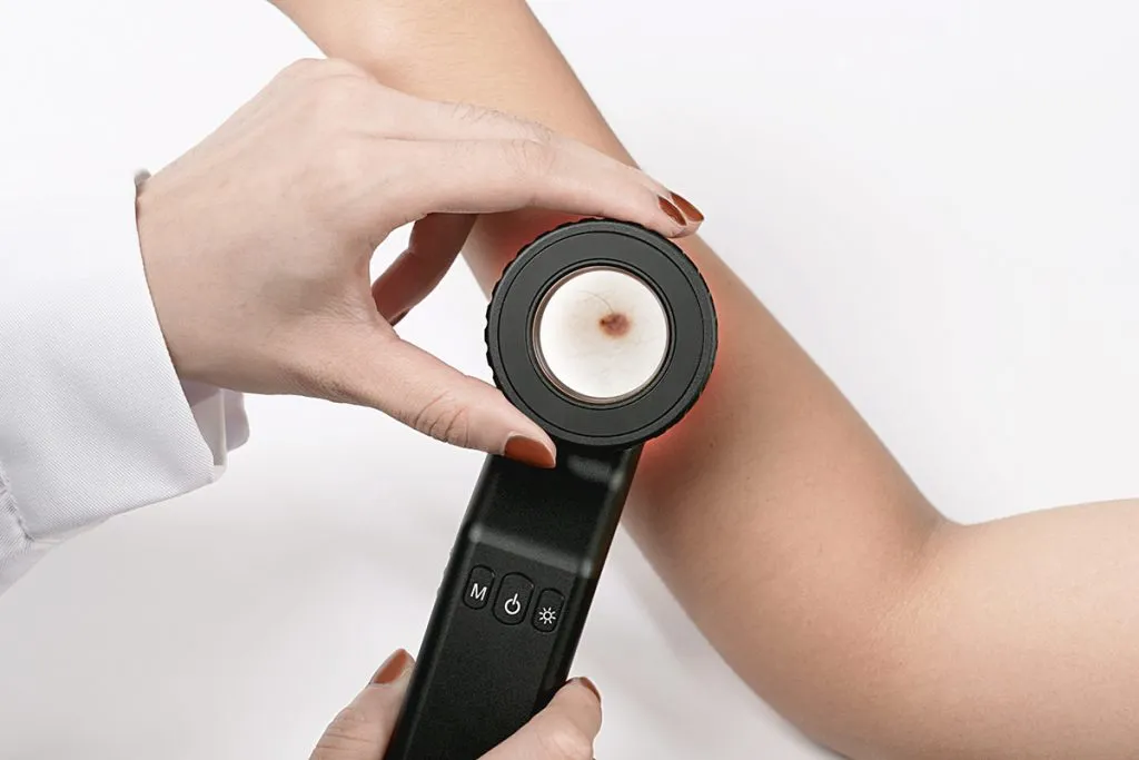
Nonpolarized dermoscope: With liquid or without liquid in the examination of skin both can work under nonpolarized dermoscope. But sometimes there is a liquid interface between the skin and device, it can enhance the visualization of subsurface by reducing the reflection from skin surface. And it commonly needs to contact the skin directly.
Polarized dermoscope: Under polarized dermoscopy, it is no requirement to use liquid interface on the skin when do examination for skin situation. And it also does not need to directly contact the skin. It brings much convenience for checking and avoids the risk of cross infection.
Amber light dermoscope: Amber light is a specialized dermoscope device for a certain features for skin. And amber light belongs to the catalogue of polarized light. So it also no needs liquid interface on the skin. It does not require to touch the skin directly.
Dermatologists will chose the suitable inspection ways to observe skin situations. Sometimes it needs to use three of them for comprehensive
information.
Comparison of dermatoscopio:price
Nonpolarized dermoscope: Compared to polarized dermoscope, nonpolarized dermoscope is more favourable.
Polarized dermoscope: Polarized dermoscope is more expensive than nonpolarized dermoscope.
Amber light dermoscope: The price of amber light dermoscope is affordable.
Commonly, a dermoscope combines the types/modes of polarized light with nonpolarized light, even amber light is also added. These two or three models can toggle mutually.
Comparison of dermatoscopio:skin conditions
Colors and structures of images under dermoscope
Nonpolarized dermoscope: Nonpolarised dermoscopy is better to exhibit peppering, milia-like cysts, blue-white colours, and comedo-like openings.
Polarized dermoscope: Polarised dermoscopy is better in displaying dermal vessels, pink/red colours, variable pigmentation, and white shiny structures.
Amber light dermoscope: Amber light dermoscope brings a enhanced and contrastive visualization of the structures for superficial skin, like nail fold capillaries.
The depth of visualised structures
Nonpolarized dermoscope: Nonpolarised dermoscopy can be used to inspect the superficial layers of skin (the superficial epidermis to the dermo-epidermal junction).
Polarized dermoscope: Polarised dermoscopy is designed to detect the deeper layers of skin (the dermo-epidermal junction and superficial dermis).
Amber light dermoscope: Amber light dermoscope highlight the visualization of superficial skin structures, especially in the superficial epidermis to the dermo-epidermal junction.
Detected skin disease types
Nonpolarized dermoscopy, polarized dermoscopy and amber light dermoscopy are three different types or models of dermoscope that helps dermatologists to detect the differences of the structures and patterns of the skin conditions by a detailed analysis. But there are still tiny differences in the diagnosis of skin disease among them.
Nonpolarized dermoscope: Nonpolarized dermoscope is a valuable device that can help to detect various skin conditions and diseases particularly in the superficial layers of skin and the superficial epidermis to the dermo-epidermal junction.
There are some common skin disease that can be better detected by a nonpolarized dermoscope, such as miliary cysts and blue-white veil. Because both milia-like cysts and blue-white veils occur in the epidermis by superficial changes therefore, they are better observed under nonpolarized dermoscope.
Polarized dermoscope: Shiny white structure is associated with an increase in collagen in the superficial layer of the dermis, so it is better observed under polarized dermoscope. Additionally, when polarized light encounters a birefringent structure, such as collagen, its polarization rapidly randomizes.Besides, blood vessels are located in the layer of dermis that is better observed under polarized dermoscope. What’s more, because polarized dermoscope without skin contact direct, lacking of the effect from pressure, vascular blush and blood vessels both can be presented with a clear and evident looks under polarized dermoscope.
Amber light dermoscope: By providing better contrast due to its wavelength of amber or yellow light, amber light dermoscope is suitable for observe the vascular changes of dermal, nail fold capillaries, and dermal pigmentation, where these structures highlight the black color against the yellow background.
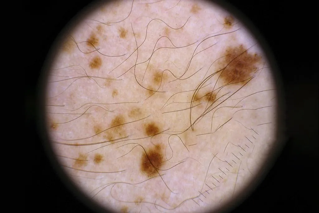
An example of a miliary cyst can be better observed under nonpolarized dermoscope.
Examples of blue and white veils are also better observed under nonpolarized dermoscope.
An example of shiny white structure can be better observed under polarized dermoscope.
Otherwise, an example of nail fold capillaries can be better observed under amber light dermoscope.
Nonpolarized light, polarized light and amber light exhibit particular patterns in prominence. Dermatologists will adopt different types of dermoscopio according to the skin conditions. The information provided by these three are complementary. It is recommended that these three all be used in clinical diagnosis. And recent dermatoscope have these three or two types together for a better visualization and better accurate diagnosis.


