


Article
Dermascope vs Dermatoscope: Exploring the Diagnostic Power beyond the Magnifying Glass
Dermascope vs Dermatoscope What is a dermatoscope?Dermatoscope, also know as dermoscope or epiluminescence microscope. It is a useful and dependable hand-held microscope device for skin examination, such as melanoma, alopecia areatap, onychosis,etc. This device allows sharp examination of skin structure by high quality magnification and super lighting system. Under dermatoscope, specific patterns and features of…
Dermascope vs Dermatoscope
What is a dermatoscope?
Dermatoscope, also know as dermoscope or epiluminescence microscope. It is a useful and dependable hand-held microscope device for skin examination, such as melanoma, alopecia areatap, onychosis,etc. This device allows sharp examination of skin structure by high quality magnification and super lighting system. Under dermatoscope, specific patterns and features of skin can be showed clearly and precisely.
Dermascope vs dermatoscope: dermoscope is the synonyms of dermatoscope.
What is the use of dermatoscope?
Dermatoscope can record the images for comparison in future. What’s more, it can monitor and assess the structure of the reticular dermal depth. The basic principle of dermatoscope is to take a translucency of the skin lesion so that it can be studied at high magnification to visualize subtle features.
Dermatoscope can magnify the outer layer of the skin greatly. Under dermatoscope, skin doctors can inspect skin colors, shapes, sizes and patterns more precisely to help distinguish and diagnose a variety of skin situations.
Skin doctors can use dermatoscope to examine situations which not related to lesions, such as melasma, hair losss, scabies, etc.
With the help of dematoscope, doctors can identify non-cancerous lesion, including dermatofibromas, warts and so on.
Dermatoscope can also help to diagnose cancerous lesions, like basal cell carcinoma, nodular melanoma, melanoma, etc.
Information from dermatoscope helps doctors to evaluate whether skin disease is progressing and whether treatment is needed or not. And it enhance doctor’s confidence in diagnosing skin situation and also greatly saves doctor’s time.
What are the types of dermatoscope?
There are many types of dermoscopes, each designed for a specific use and function. Here are the key types:
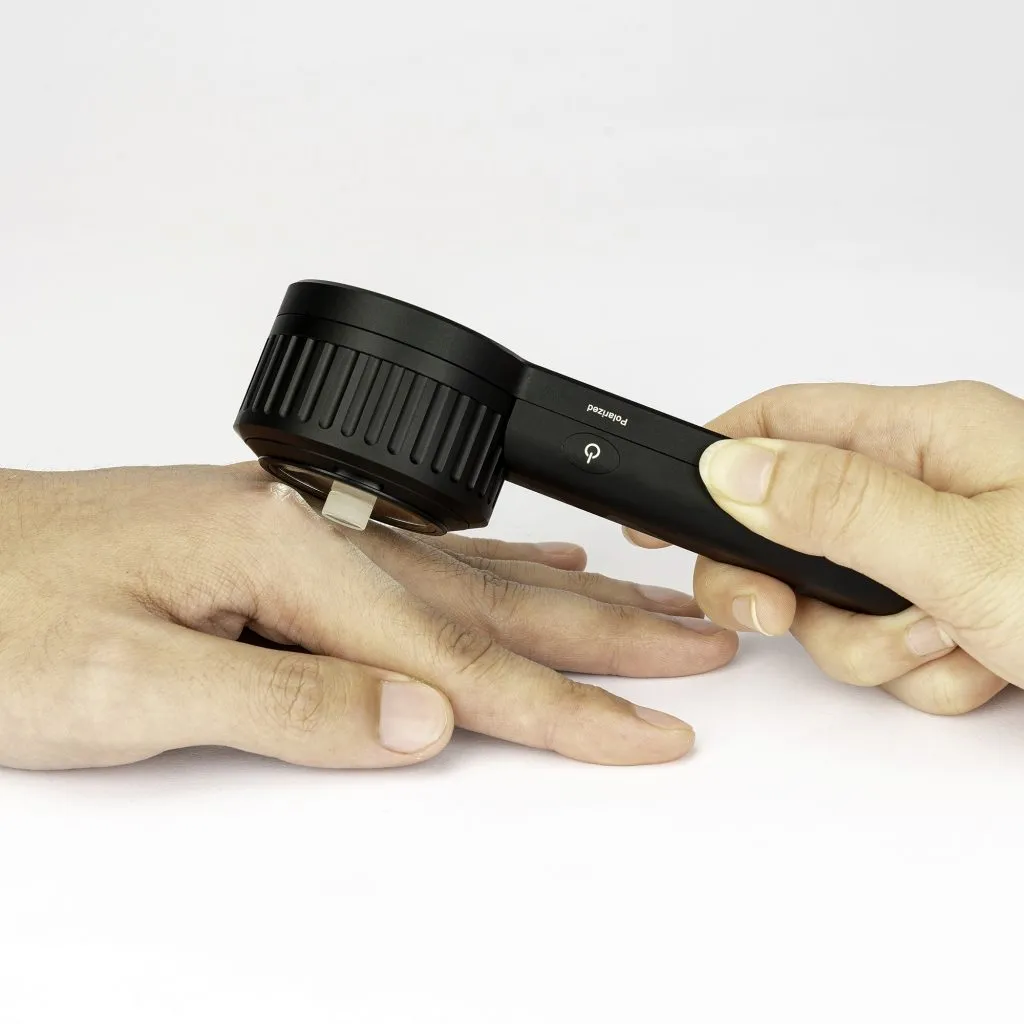
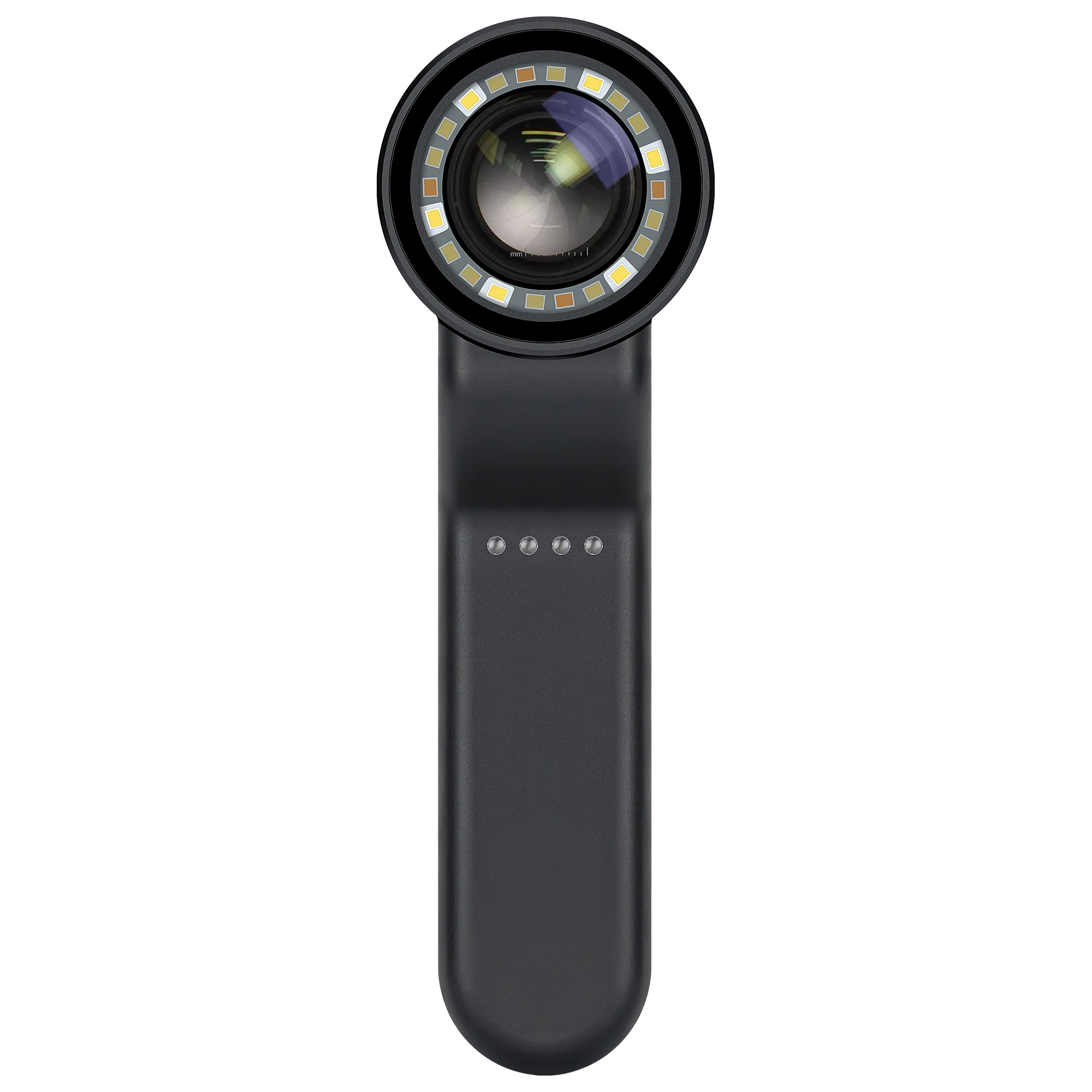
• Hand-held portable dermatoscope: This is a common type and it is often used due to its convenience and mobility. It typically includes a transparent light source and an amplifying optical device, usually providing a magnification of about 10-fold.
• Stationary Mounted Type: This kind of dermatscope is designed to capture whole body images in one shot. They are typically integrated with image analysis algorithms to generate a three-dimensional model of the human body and use artificial intelligence to label and analyze lesions.
In addition, dermatoscope have different models of operation, such as unpolarized light contact, polarlized light contact, polarlized non-contact, etc.
• Unpolarized light, contact: Unpolarized light is a natural light with intrinsically incoherent and it has an electric field that oscillates in all directions. It can provides information for superficial skin.
• Polarized light, contact: Polarized light is intrinsically coherenn and it has an electric field that oscillates in only one direction. Polarlized light contact can reveal the depth of skin structures.
• Polarized light, non-contact: As its name, skin can be inspected without contact. It is very suitable especially in sensitive area. By using polarized light, it can eliminate the reflection and surface glare of skin to show the dermis structures of skin.
What are the clinical effects of dermatoscope?
In the field of dermatology, the clinical effects of using a dermatoscope is of great significance. Especially in the monitoring and diagnosis of skin lesions. Here are some of the main advantages:
• Enhanced Diagnosis: Dermatoscopy helps dermatologists to identify pigmented skin lesions more accurately, which can avoid some unnecessary surgery or skin biopsies.
• Monitoring Changes: Dermatoscope helps dermatologists to detect moles and other pigmented skin lesions over time for any changes, which is very important for early monitoring of skin cancer.
• Non-Invasive: The whole procedure is painless and non-invasive, and it is suitable for all skin types and ages.
• Visualization of Subsurface Structures: Dermatoscopy offers a amplifying view of the dermoepidermal junction, epidermis, and papillary dermis, which can not be saw by the naked eye.
• Digital Documentation: Images captured by the dermatoscope can be digitally recorded for sequential monitoring or storage,helping to observe suspicious lesions carefully.
These clinical effects contribute to the early examination and treatment of skin cancers such as basal cell carcinoma and melanoma, as well as in the treatment of other skin diseases.
Detection of melanoma with dermatoscope
Dermatoscopy is a noninvasive, aided instrument by dermologists used for the examination of various skin lesions, like melanoma. Dermascope vs dermatoscope: Dermoscopy is performed with a handheld device called a dermatoscope.
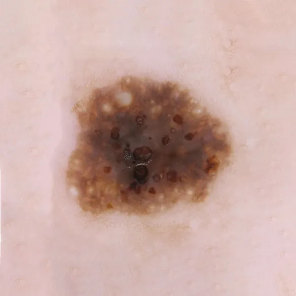
The detection of melanoma with dermatoscope is a critical process that involves identifying specific patterns and structures that are indicative of the disease. Here are some key points regarding melanoma detection with a dermatoscope, such as early detection, specific features, improved accuracy.
What’s another name of dermatoscope?
A dermatoscope is also known by several other names, including: deromscope, dermoscopy, dermatoscopy, Epiluminescence Microscopy, Epiluminescence Microscopy, Incident Light Microscopy and so on. Dermascope vs dermatoscope, deroscope is another name of dermatoscope.
Dermatoscope vs Magnifying Glass
Do dermatologists use magnifying glass?
Yes, for a more general examination of the skin, dermatologists may use simple magnifying glass to inspect the skin. And a magnifying glass is usually used to show a magnified image of objects.
Dermatoscope vs magnifying glass:While a dermatoscope is a specialized tool that connects physic and optic. It can reveal the dermis and epidermis of skin clearly through eliminate the light glare and reflection of skin.Dermascope vs dermatoscope: Dermoscope is also know as dermatoscope, is a valuable and dependable device for dermatologists.
What is the difference between dermatoscope and magnification?
Dermatoscope vs magnifying glass:Dermatoscope (Dermoscope) is a valuable aid device used by skin doctors to examine skin lesions with more details. It combines illumination with magnification to release a clear view of underground structures that are invisible to the routine examination by naked eye. Dermatoscope vs magnifying glass, here’s the differences as below:
• Magnification: Usually it means to use lenses or digital technology to enlarge an image. It does not contain a light source, nor does it display subsurface of the skin in details.
• Dermoscopy: While dermoscopy is working, it magnifies the surface of the skin, at the some time, it also illuminates the skin with specific types of light. So it greatly enhance the view of skin structures and patterns under the surface, which is essential for diagnosing skin diseases such as melanoma.
In essence,Dermatoscope vs magnifying glass, magnification can amplify objects and make it looks larger. But the function of dermoscopy is far more than this. It can provide a more comprehensive view by combining magnification and illumination, helping skin doctors to diagnose more accurately.
What Is a Dermatoscope? Handheld Dermascope vs Dermatoscope for Dermatologist | IBOOLO
Discover what is a dermatoscope vs a dermascope for dermatologist use. Compare a handheld dermatoscope vs a magnifying glass and explore IBOOLO’s advanced tools for precise skin analysis.
Dermascope vs Dermatoscope: Magnification and Uses Beyond the Magnifying Glass
Understanding the differences between a dermascope vs dermatoscope, as well as dermatoscope vs magnifying glass, is crucial for dermatologists and individuals monitoring skin health. IBOOLO’s advanced handheld dermatoscopes, like the DE-3100 and DE-4100, offer superior dermoscopy magnification and lighting, surpassing traditional magnifying glasses. This guide explores their diagnostic power, dermatoscope uses, and why they’re essential tools for dermatology, helping you choose the right device for precise skin analysis.
What Is a Dermatoscope?
A dermatoscope, also known as a dermoscope or epiluminescence microscope, is a handheld device designed for detailed skin examination. Unlike a magnifying glass, a dermatoscope combines high-quality optics with specialized lighting (polarized or non-polarized) to reveal subsurface skin structures, such as pigmentation and vascular patterns. IBOOLO’s dermatoscopes provide 10x dermatoscope magnification, ideal for diagnosing conditions like melanoma, alopecia areata, and onychosis. The terms dermascope or dermatoscope are often used interchangeably, though “dermatoscope” is the standard medical term, synonymous with dermoscope for dermatologist use.
Dermascope vs Dermatoscope: Are They Different?
The debate of dermascope vs dermatoscope often arises due to terminology. In essence, a dermascope is the same as a dermatoscope—both refer to a handheld device used for dermoscopy. The term “dermoscopy” encompasses skin surface microscopy, also called epiluminescence microscopy or incident light microscopy. IBOOLO’s dermatoscopes, like the DE-3100, use advanced optics to provide clear visuals, whether labeled as a dermascope or dermatoscope. The key is their ability to offer precise dermoscopy magnification, typically 10x, for professional and personal skin monitoring, always in conjunction with medical advice.
Dermatoscope vs Magnifying Glass: Key Differences
Comparing a dermatoscope vs magnifying glass highlights significant differences in functionality:
- Magnification Power: A dermatoscope offers 10–100x dermoscopy magnification, compared to a magnifying glass’s 3–8x, revealing deeper skin structures like the dermis and epidermis.
- Lighting Technology: Dermatoscopes use polarized and non-polarized light to eliminate glare, unlike magnifying glasses, which rely on ambient light and cause reflections.
- Diagnostic Precision: Dermatoscopes enhance diagnostic accuracy by up to 35% for melanoma detection, while magnifying glasses provide only surface-level views. [](https://www.medicalnewstoday.com/articles/dermatoscope)
IBOOLO’s handheld dermatoscopes, with features like polarized light and smartphone compatibility, far surpass magnifying glasses for detailed skin analysis, making them a preferred dermoscope for dermatologists.
Handheld Dermatoscope vs Magnifying Glass
A handheld dermatoscope vs magnifying glass comparison emphasizes portability and precision. Handheld dermatoscopes, such as IBOOLO’s DE-3100 (180g) and DE-4100 (325g), offer:
- Portability: Lightweight and battery-powered, ideal for clinical or home use.
- Advanced Optics: 10x dermatoscope magnification with multiple lighting modes (polarized, non-polarized, amber) for versatile skin examination.
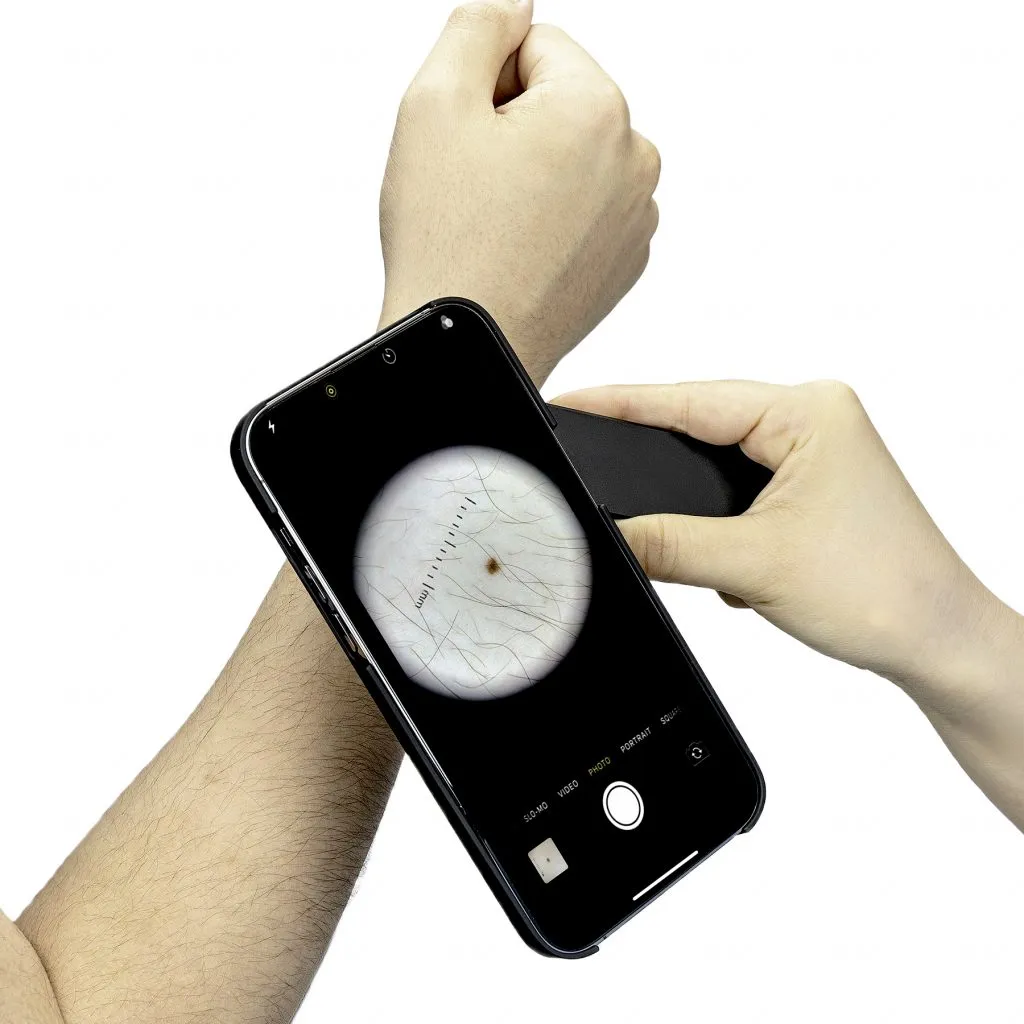
- Image Capture: Smartphone adapters enable photo documentation, unlike magnifying glasses, which lack this capability.
These features make handheld dermatoscopes superior for diagnosing conditions like basal cell carcinoma or monitoring treatment progress, far beyond the capabilities of a magnifying glass.
Dermatoscope Uses in Dermatology
Dermatoscope uses extend across various dermatological applications, making them indispensable for skin health management:
- Skin Cancer Detection: Enhances early identification of melanoma, basal cell carcinoma, and squamous cell carcinoma with up to 92% sensitivity. [](https://www.medicalnewstoday.com/articles/dermatoscope)
- Hair and Scalp Analysis: Diagnoses conditions like alopecia areata or tinea capitis by examining scalp patterns.
- Nail Disorders: Identifies onychosis or nailfold capillaroscopy changes for systemic disease detection.
- Infectious Diseases: Detects scabies burrows or fungal infections with precision. [](https://en.wikipedia.org/wiki/Dermatoscopy)
IBOOLO’s dermatoscopes support these applications with high-resolution imaging, ensuring accurate observation when paired with professional medical evaluation.
Dermoscopy Magnification and Its Importance
Dermoscopy magnification is a critical feature of dermatoscopes, typically ranging from 10x to 100x, depending on the device. Handheld dermatoscopes like IBOOLO’s DE-3100 provide 10x magnification, sufficient for most clinical needs, while video dermatoscopes can reach 70–100x for detailed research. This magnification allows visualization of:
[](https://www.medicalnewstoday.com/articles/dermatoscope)- Pigment Networks: Irregular patterns indicating potential malignancy.
- Vascular Structures: Atypical blood vessels associated with skin cancers.
- Surface Features: Milia-like cysts or comedo-like openings in benign lesions.
Higher dermatoscope magnification enhances diagnostic precision, making IBOOLO’s devices ideal for both dermatologists and trained individuals monitoring skin changes.
Why Choose a Dermatoscope for Dermatology?
A dermoscope for dermatologist use is a vital tool, often compared to a stethoscope for its diagnostic value. Benefits include:
[](https://abcnews.go.com/Health/detecting-skin-cancer-handheld-magnifying-tool-naked-eye/story?id=59905561)- Non-Invasive Diagnosis: Reduces unnecessary biopsies by distinguishing benign from malignant lesions.
- Improved Accuracy: Increases melanoma detection sensitivity to 92% compared to 76% with naked-eye exams. [](https://www.medicalnewstoday.com/articles/dermatoscope)
- Versatile Applications: Supports diagnosis of pigmented lesions, inflammatory conditions, and hair/scalp disorders.
IBOOLO’s dermatoscopes, with features like polarized light and smartphone integration, empower dermatologists to deliver precise, patient-centered care.
IBOOLO’s Dermatoscope Features
IBOOLO, a leading manufacturer since 2012, offers dermatoscopes designed for professional and personal use:
- DE-3100: Lightweight (180g), 10x magnification, polarized/non-polarized light, smartphone-compatible.
- DE-4100: Robust (325g), premium optics, multiple lighting modes for detailed skin analysis.
- Smartphone Integration: Universal adapters for image capture on iPhones, Androids, or cameras, enhancing teledermatology.
These features make IBOOLO’s dermatoscopes a top choice for dermascope or dermatoscope needs, offering unmatched clarity and convenience.
Future of Dermatoscope Technology
Advancements in dermatoscope technology are shaping the future of dermatology:
- AI Integration: Enhances automated lesion analysis for faster, more accurate diagnoses.
- Teledermatology: Supports remote consultations via 5G and cloud-based image sharing. [](https://www.nature.com/articles/s41746-022-00587-9)
- Higher Magnification: Video dermatoscopes with 20–100x magnification for research and detailed analysis.
IBOOLO is at the forefront, developing dermatoscopes with AI compatibility and telemedicine features to improve access to skin care.
FAQs About Dermascope vs Dermatoscope
What is a dermatoscope?
A dermatoscope is a handheld device that combines 10x magnification and specialized lighting to examine skin lesions, used for diagnosing melanoma, alopecia, and other conditions.
Is a dermascope different from a dermatoscope?
No, dermascope and dermatoscope are synonymous terms for the same device used in dermoscopy, offering advanced magnification and lighting for skin analysis.
How does dermatoscope magnification compare to a magnifying glass?
Dermatoscope magnification (10–100x) far exceeds that of a magnifying glass (3–8x), providing clearer views of subsurface skin structures for accurate diagnosis.
What are the main dermatoscope uses?
Dermatoscopes are used for detecting skin cancers, hair/scalp disorders, nail conditions, and infectious diseases like scabies, enhancing diagnostic precision.
How does dermatoscope magnification compare to a magnifying glass?
Dermatoscope magnification, ranging from 10–100x, far exceeds the 3–8x of a magnifying glass, providing clearer views of subsurface skin structures for accurate diagnosis. IBOOLO’s handheld dermatoscopes, like the DE-3100, deliver 10x magnification, ideal for dermatologist use in identifying melanoma or other conditions.
What are the main dermatoscope uses?
Dermatoscope uses include detecting skin cancers, hair/scalp disorders, nail conditions, and infectious diseases like scabies, enhancing diagnostic precision. IBOOLO’s dermatoscopes support these applications with high-resolution imaging, making them a top dermoscope for dermatologist needs.
Dermatoscope for Dermatology: A Must-Have Tool
A dermoscope for dermatologist use is indispensable, often compared to a stethoscope for its diagnostic value. Unlike a magnifying glass, a handheld dermatoscope offers advanced dermoscopy magnification and lighting, enabling non-invasive diagnosis of conditions like basal cell carcinoma, squamous cell carcinoma, and inflammatory diseases. IBOOLO’s dermatoscopes, with features like polarized light and smartphone compatibility, empower dermatologists to monitor skin changes with precision, reducing unnecessary biopsies and improving patient outcomes. Studies show dermoscopy magnification enhances melanoma detection sensitivity to 92%, compared to 76% with naked-eye exams, making it a cornerstone of modern dermatology.
IBOOLO’s Handheld Dermatoscope Features
IBOOLO, a trusted manufacturer since 2012, offers dermatoscopes optimized for both professional and personal use. The debate of dermascope or dermatoscope is irrelevant with IBOOLO’s devices, as they deliver consistent performance under either label. Key features include:
- DE-3100: Lightweight (180g), 10x dermatoscope magnification, with polarized and non-polarized light for versatile skin analysis.
- DE-4100: Robust (325g), premium optics, and multiple lighting modes, ideal for detailed dermatology diagnostics.
- Smartphone Integration: Universal adapters for iPhones, Androids, or cameras, enabling image capture for teledermatology and documentation.
These features make IBOOLO’s handheld dermatoscopes a top choice for dermatologist use, surpassing traditional glass magnifiers in functionality and convenience.
Future Trends in Dermatoscope Technology
The future of dermascope vs dermatoscope technology is evolving rapidly, enhancing their role as a dermoscope for dermatologist applications:
- AI Integration: Automates lesion analysis, improving diagnostic accuracy for conditions like melanoma.
- Teledermatology: Supports remote consultations via 5G and cloud-based image sharing, expanding access to skin care.
- Enhanced Magnification: Video dermatoscopes with 20–100x dermoscopy magnification for research and detailed analysis.
IBOOLO is leading these advancements, offering dermatoscopes with AI compatibility and telemedicine features, ensuring they remain a preferred dermatoscope for dermatology.
Why Choose IBOOLO for Dermascope or Dermatoscope Needs?
Whether you’re debating dermascope vs dermatoscope or comparing a handheld dermatoscope vs magnifying glass, IBOOLO’s devices stand out for their precision and versatility. Designed for dermatologist use, models like the DE-3100 and DE-4100 offer 10x dermatoscope magnification, multiple lighting modes, and smartphone integration, making them ideal for both clinical and personal skin monitoring. Unlike a basic glass magnifier, IBOOLO’s dermatoscopes provide detailed visuals of skin structures, supporting early detection and ongoing management of skin conditions when used as an auxiliary tool alongside professional medical advice.
Dermatoscope as the Future of Skin Analysis
Understanding what is a dermatoscope and its advantages over a magnifying glass highlights its critical role in dermatology. The comparison of dermascope vs dermatoscope reveals they are synonymous, with IBOOLO’s handheld dermatoscopes offering unmatched dermoscopy magnification and dermatoscope uses. From detecting skin cancers to analyzing hair and nail disorders, these devices empower dermatologists and individuals to monitor skin health with precision. Choose IBOOLO’s advanced dermatoscopes for a reliable, non-invasive solution that transcends traditional glass magnifiers, ensuring accurate skin analysis for dermatologist needs.
Recommended reading
High Quality Dermoscopy Meaning Created in Our Products Supply Based in China - IBOOLO
Our China products supply hub couples world-class portability with elite precision, using seasoned expertise to develop high quality dermoscopy meaning for flawless skin visualization anywhere through compact size.
China Skin Cancer Dermoscopy Products Supply Specializes in Professional Items - IBOOLO
Our China products supply creates clinical quality Professional skin cancer dermoscopys enabling powerful skin magnification from anywhere through thoughtful craftsmanship.
China Products Supply Provides Wholesale Dermatoscope Phone Attachments for Clients - IBOOLO
As an expert China products supply, we use exacting wholesale production methods to manufacture high-quality dermatoscope phone attachment solutions tailored for every customer.


