


Article
Dermatologist Magnifying Glass
There are several kinds of dermatological magnification tools and the following are some of the common ones together with their uses. Handheld magnifiers are the most basic and are for limited use on small areas remain of free skin. and Head are mounted useful magnifiers for are examining worn and on manipulating the the head…
There are several kinds of dermatological magnification tools and the following are some of the common ones together with their uses. Handheld magnifiers are the most basic and are for limited use on small areas remain of free skin. and Head are mounted useful magnifiers for are examining worn and on manipulating the the head skin such in that detail. the Digital hands magnifiers of are the instruments doctor that use electronic means of enhancing the size of an object for observation and the enhanced image is displayed on an electronic screen.
The dermatoscope is a medical instrument used by dermatologists to examine skin lesions and is one of the most popular magnifying observation devices in dermatology. Other types of dermoscope include polarized dermoscope that can take away scattered light from the skin surface, thus enabling doctors to have a clear view of the internal structure of skin lesions.
Technical Characteristics of Different Types of Dermatoscopes
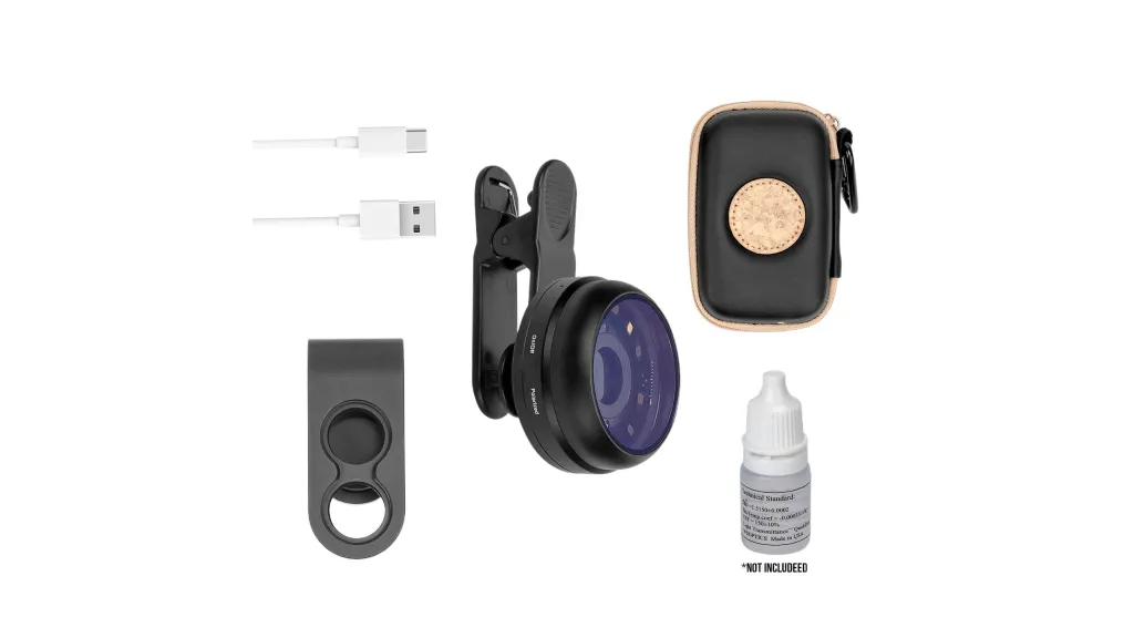
IBOOLO optical dermatoscopes are categorised into handheld dermatoscopes and pocket dermatoscopes. Pocket dermatoscopes can be regarded as simple versions of dermatoscopes. IBOOLO DE-200 and DE-300 have a magnification of 6X and a field of view of 12mm. DE-200 is the first generation of dermatoscopes from IBOOLO, which has only polarised light. DE-300 is an upgraded version, which has both polarised and unpolarised light. DE-400 is the newest version of IBOOLO’s pocket dermatoscopes at present. DE-400 is IBOOLO’s newest pocket dermatoscope with 10X magnification and 20mm window.
The DE-400 is the latest IBOOLO pocket dermatoscope with 10X magnification and a 20mm window, both polarised and non-polarised.
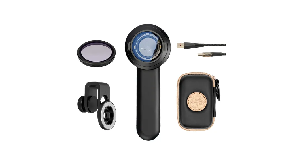
IBOOLO’s handheld dermatoscopes are currently available in two models, the DE-3100 and DE-4100, both of which have the same 10X magnification, with the difference that the window of the DE-3100 is 32mm and that of the DE-4100 is a bit larger at 48mm. the illumination systems of both models are more complete and versatile than those of the Pocket Dermatoscope. They have four lighting modes, white light polarised, amber light, mixed light polarised and white light unpolarised, which are suitable for the observation of a wide range of skin lesions.
The whole series of IBOOLO dermatoscopes basically adopts the all-metal appearance and aluminium alloy manufacture. The metal feature can effectively prevent the dermatoscope from being damaged by dropping or external impact, and improve the durability.
Dermoscopy in Clinical Diagnosis
Dermoscopy is most extensively used in the day to day clinical practice for the purpose of skin lesions’ detailed observation. The pigmented lesions of the skin, including moles, freckles and pigmented spots can be seen in dermoscopy that cannot be seen with the naked eye. It is also able to see the tiny structures underneath the skin surface which is vital in the diagnosis of certain lesions. The early detection of skin cancer particularly melanoma cannot be done without dermoscopy in its early stages. Melanoma is generally an irregularly shaped lesion with an irregular color pattern and poorly defined edges. Through dermoscopy with its magnification and high resolution imaging, doctors are able to identify these early signs such as the pigmented glaze and asymmetrical vascular patterns and hence decide whether the lesion is dangerous or not. Dermoscopy is not only used for the examination of skin surface but can also be employed in the diagnosis of hair and scalp disorders. The dermoscopy of the scalp offers a clear view of the density of the hair, the state of the hair follicles and any abnormal skin lesions such as psoriasis and tinea capitis. In addition, dermoscopy is particularly useful in the evaluation of nail diseases as it provides a clear view of the nail plate, color and structures as well as lesions beneath the nail. For instance, dermoscopy can help in the identification of onychomycosis, nail injuries, peronychia, and other vascular abnormalities under the nails.
How to Choose the Right Dermatologist Magnifying Glass
If you want to buy IBOOLO dermatoscope and don’t know how to choose, then you can read this board carefully.
If your budget is not high, then you can first consider our pocket dermatoscope series. Each of the Pocket Skin Mirrors has a polarised light feature that will satisfy your basic use of skin mirrors. However, the Pocket Dermoscopes range can only be connected to a mobile phone for skin examination and have low magnification except for the DE-400.
If you have a relatively high budget, then consider the IBOOLO handheld dermatoscope series. The handheld dermatoscopes have a magnification of 10X, which can help doctors to observe many details that are not visible to the naked eye. It also has a variety of light modes to help the doctor easily observe all types of lesions. However, the price is higher for beginners.
Tips for the Proper Use of Dermatologist Magnifying Glass
When skin magnification is performed, the magnification of the dermatoscope is adjusted according to the size of the examined area and the details required. Observe the window of the dermatoscope until the area under observation is clear. As well as to avoid too close contact between the dermatoscope and the skin, so as not to affect the observation effect or produce errors.
After use, be sure to clean the lens of the dermatoscope to avoid oil, dust, etc. affecting the image quality. Use special lens cleaning paper or a fibre-free cleaning cloth to gently wipe the surface of the lens. At the same time, this operation can avoid cross-infection between patients to the greatest extent possible.
Through non-invasive examination of dermoscopy, a lot of information can be obtained, but the final diagnosis may still be confirmed by pathology and skin biopsy. For suspicious lesions found during dermoscopy, doctors may consider further pathological section examination.
Dermatologist Magnifying Glass in Modern Dermatological Practice
Dermoscopy has become an indispensable tool in modern dermatological clinical diagnostics, especially in the early screening of skin lesions, the diagnosis of skin cancer, and the precise evaluation of skin diseases. Moreover, dermoscopy reduces skin irritation and discomfort while avoiding patient anxiety and pain associated with biopsies.
Dermatologist Magnifying Glass 2025: Professional Buying Guide & Comparison | IBOOLO
Discover how to choose the best dermatology magnifying lens for accurate skin diagnosis. Compare top brands, key features, and expert tips for clinical or home use.
2025 Dermatologist Magnifying Glass Buying Guide: Accurate Diagnosis Starts With Choosing The Right Tool
Choosing the right dermatology magnifying lens is the first step toward professional-grade skin examination. This guide cuts through the complexity to help dermatologists, medical professionals, and skincare enthusiasts select the perfect tool for precise diagnosis and effective patient care.
How to Choose Your Dermatologist Magnifying Glass: 5 Key Buying Factors
Selecting the right dermatology magnifying lens requires careful consideration of several technical and practical factors. The ideal tool balances optical clarity, illumination, and ergonomics for your specific use case.
Key Factors for Selection:
- Magnification Power (10x-20x): 10x magnification is standard for general examination, while 15x-20x provides enhanced detail for specialized diagnosis.
- Illumination Type (LED vs. Halogen): LED offers cooler operation and better energy efficiency, while halogen provides warmer light with potentially better color rendering.
- Lighting Modes (Polarized/Non-Polarized): Polarized light reduces surface glare to reveal subsurface structures, while non-polarized light enhances surface texture details.
- Ergonomics & Weight: Balanced design and lightweight materials (200-400g) prevent hand fatigue during extended examinations.
- Optical Quality: Multi-coated, anti-reflective lenses with minimal distortion are essential for accurate diagnosis.
Dermatologist Magnifying Glass vs. Dermatoscope: Core Differences & Selection Advice
While both tools serve examination purposes, understanding their fundamental differences ensures you select the appropriate instrument for your clinical needs and budget.
Comparison Analysis:
- Technology: Dermatologist magnifying glasses provide optical magnification with basic illumination, while dermatoscopes incorporate advanced lighting systems and often digital capabilities.
- Cost Factor: Professional magnifying glasses range from $150-$500, whereas dermatoscopes typically start at $500 and can exceed $2000 for digital systems.
- Portability: Magnifying glasses offer superior portability for field practice or multiple examination rooms.
- Diagnostic Capability: Dermatoscopes provide more advanced features for pigment pattern analysis and documentation.
Top Dermatology Magnifying Lens Brands & Models: In-Depth Comparison
The market offers several reputable brands producing professional-grade dermatology magnifying lenses. This comparison highlights key products based on performance, features, and user feedback.
Leading Brands Overview:
- IBOOLO Professional Series: Offers excellent value with 10x/20x magnification options, dual LED illumination, and lightweight design (280g).
- DermLite Dermatoscopes: Industry leader with multiple magnification options and patented polarization technology.
- Heine Dermatoscopes: German-engineered optics with exceptional clarity and rugged construction.
- Welch Allyn: Trusted medical brand offering reliable basic models for general practice.
Essential Applications of Dermatology Magnifying Lenses in Clinical Practice
Dermatologist magnifying glasses serve multiple critical functions in clinical settings, enhancing diagnostic accuracy across various skin conditions.
Primary Clinical Applications:
- Skin Cancer Screening: Early detection of melanoma, BCC, and SCC through detailed examination of lesion borders, color patterns, and structures.
- Inflammatory Condition Assessment: Close monitoring of psoriasis, eczema, and rosacea progression and treatment effectiveness.
- Pigment Disorder Evaluation: Detailed analysis of melasma, vitiligo, and other dyschromias for accurate diagnosis and treatment planning.
- Procedural Guidance: Enhanced visualization during cosmetic procedures, biopsies, and lesion removals.
Technical Specifications: Understanding Dermatology Magnifying Lens Capabilities
The performance of a dermatologist magnifying glass depends on specific technical parameters that determine its diagnostic utility.
Critical Specifications:
- Field of View: Ranges from 10mm to 40mm depending on magnification power.
- Working Distance: Typically 2-4 inches for comfortable examination without instrument contact.
- Light Source Intensity: LED systems providing 3,000-10,000 lux illumination for optimal visualization.
- Battery Life: Rechargeable systems offering 2-8 hours of continuous operation.
Maintenance and Care for Your Dermatologist Magnifying Glass
Proper maintenance ensures optimal performance and longevity of your dermatology magnifying lens investment.
Essential Maintenance Practices:
- Clean lenses regularly with microfiber cloth and approved optical cleaning solution
- Disinfect housing and contact surfaces between patients with appropriate medical-grade disinfectants
- Store in protective case when not in use to prevent scratches and damage
- Regularly check and maintain battery systems for cordless models
- Schedule professional calibration and servicing for high-end models annually
Frequently Asked Questions: Dermatologist Magnifying Glasses
What magnification power is best for general dermatology practice?
10x magnification is considered the standard for most general dermatology applications, providing sufficient detail for routine examinations while maintaining a good field of view.
Can I use a dermatology magnifying lens for self-examination?
While professionals should perform definitive diagnoses, high-quality magnifying glasses can help individuals monitor existing lesions and identify changes that warrant professional evaluation.
How important is polarized light in dermatologist magnifying glasses?
Polarized light is valuable for reducing surface glare and examining subsurface structures, particularly for pigmented lesions and vascular conditions.
What warranty should I expect with a professional-grade magnifying glass?
Reputable manufacturers typically offer 2-3 year warranties on professional instruments, covering defects in materials and workmanship.
Where can I purchase authentic dermatologist magnifying glasses?
Purchase directly from manufacturer websites, authorized medical equipment distributors, or reputable medical supply companies to ensure genuine products and valid warranties.


