Article

Dermatoscope for Dermatology

Dermoscopy, also known as epidermal transillumination microscopy, is a non-invasive, fast and easy means of examining the skin, which has a wide range of applications and plays an important role especially in different areas of dermatology. Dermoscopy has significant advantages in the diagnosis of skin tumours. By observing the pigmentation, blood vessels, texture and other…

Dermoscopy, also known as epidermal transillumination microscopy, is a non-invasive, fast and easy means of examining the skin, which has a wide range of applications and plays an important role especially in different areas of dermatology. Dermoscopy has significant advantages in the diagnosis of skin tumours. By observing the pigmentation, blood vessels, texture and other features of the skin surface, doctors can make preliminary judgement on the goodness or badness of the tumour; by analysing the inflammatory manifestations of the skin such as erythema, oedema, and pustules through the dermoscopy, doctors can diagnose a wide range of inflammatory skin diseases, such as psoriasis, eczema, and so on. Dermoscopy can also assist in the observation and diagnosis of hair diseases, nail diseases and vascular diseases.

DE-4100 Dermatoscope with Eye Piece


The Role of Dermoscopy in the Diagnosis of Skin Cancer
Melanoma is a highly malignant tumour and early detection and treatment is essential to improve patient survival. By magnifying the surface of the skin 10 to 20 times, dermoscopy is able to clearly show the fine structures of melanoma, such as irregular pigment networks or abnormal blood vessel patterns. These features are important in differentiating melanoma from other benign lesions.
Non-melanoma skin cancers mainly include the types of squamous cell carcinoma and basal cell carcinoma. These skin cancers usually show symptoms such as gradual enlargement of skin lesions, rough or scaly surface, uneven colour, bleeding or discharge, and painless ulcers.
Dermoscopy can assist the doctor in determining the benign or malignant nature of the lesion. Malignant lesions usually have abnormal morphological features, such as cellular anisotropy and increased nuclear division, whereas benign lesions usually have good cellular differentiation and cellular morphology similar to that of normal tissue.

Dermoscopy in the Diagnosis


Dermoscopy in Inflammatory Skin Diseases
Psoriasis is a common inflammatory skin disease, which is characterised dermoscopically by evenly distributed punctate, globular, circumscribed or hairpin-like blood vessels on a bright red background with diffuse white scales. Eczema is an inflammatory skin reaction with intense itching caused by a variety of internal and external factors. Under dermoscopy, the features of eczema include small flakes or fine dots of bleeding, follicular papules, oozing, and crusting.
Dermoscopy can also be used to diagnose a variety of other inflammatory skin conditions, such as pityriasis rosea and lichen planus. Pityriasis rosea presents with peripheral white scales on a yellow background (collar sign) and clustered distribution of punctate blood vessels. Lichen planus shows pearly white, yellow or bluish-white Wickham’s stripe, which may be reticular in form.


The Role of Dermoscopy in the Diagnosis of Vascular Lesions
For specific types of vascular tumours, such as spider nevus or capillary dilatation, there are typical morphological features in dermoscopy. For example, a spider nevus may present dermoscopically with a small red spot in the centre, surrounded by many tiny red blood filaments radiating in the shape of a spider’s web. Through dermoscopy, the doctor can also clearly observe the morphology and distribution of capillaries, so as to assess the degree and extent of capillary dilatation.
Dermoscopy also plays an important role in the differential diagnosis of vascular tumours. Vascular tumours include various types, such as hemangiomas and angiofibromas. Angiofibromas appear dermoscopically as nodules or masses on the skin surface with well-defined borders.

The Role of Dermoscopy in the Diagnosis of Vascular Lesions


The Use of Dermoscopy in Dermatological Treatments
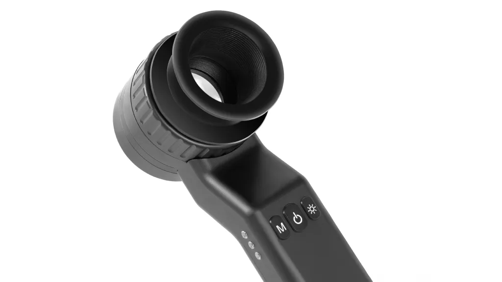
IBOOLO, as a professional dermatoscope brand, has launched different series of dermatoscopes to meet the requirements of different groups of people, and DE-3100 and DE-4100, as the high-end optical dermatoscopes of IBOOLO, play an important role in dermatological treatments, especially for the evaluation and planning of the treatment results.
IBOOLO dermatoscopes can reflect the colour and structural characteristics of the epidermis and dermal papillary layer by means of the optical magnification principle, thus assisting in the diagnosis of a wide range of skin diseases. By urging patients to have regular dermoscopy examinations, doctors can achieve dynamic follow-up of skin damage. In this process, doctors can readily determine the effectiveness of treatment and adjust the treatment plan according to changes in the condition. For example, in chloasma treatment, a skin image analysis system can be used to analyse the dermoscopic images and calculate the degree of regression and colour change of the pigmented area after treatment to determine the efficacy of the treatment.

Dermatoscope for Dermatology


Standardised Procedure for Dermoscopy
If you would like to use the IBOOLO Dermatoscope to perform a dermatoscopic examination, then take a look at the tutorial below.
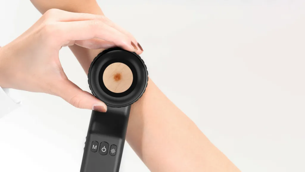
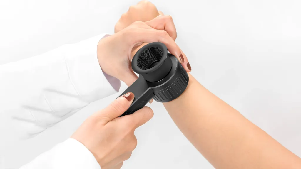
When choosing an IBOOLO dermatoscope for a full-body skin examination, first clean the surface of the skin to be examined, removing oil, make-up, and other reflective substances. Disinfect the dermatoscope lens and other examination instruments to ensure sterility. Beginning with the head, gradually work your way down to examine the skin of the entire body, noting the condition of the skin in each area. For large skin areas, a zonal examination method can be used to avoid missing them. If a dermoscopic examination of a specific area is desired, the process is the same.
Another major point of interest during the examination is focusing and choosing the distance at which to look. both the DE-3100 and DE-4100 dermatoscopes can be used either close to the skin or at a distance from the area under observation. For patients with ulcerated or inflamed lesions, the latter may be more appropriate. This provides maximum comfort for the patient.
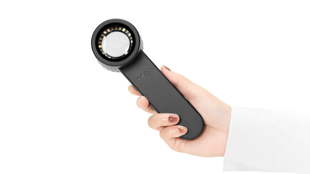
Both the DE-3100 and DE-4100 models can be connected to a mobile phone, which makes it possible to save dermatoscopic images with the mobile phone camera. By comparing dermatoscopic images on a regular basis, the doctor can visualise the changes in the lesions and make a more rational treatment plan.


Dermatoscopy: Your Guide to Skin Health Monitoring - IBOOLO

Explore IBOOLO dermatoscopy, a non-invasive tool that reveals skin details beyond the naked eye. Discover dermoscopy applications, from identifying moles to diagnosing skin conditions, and learn how it aids in early detection with professional guidance.

Dermatoscopy: Your Optical Ally for Skin Health

When you notice a new mole or a change in an existing skin lesion, uncertainty can arise. Dermatoscopy, also known as dermoscopy, serves as a powerful, non-invasive tool to bridge the gap between visual limitations and health concerns. This advanced optical technique acts like a magnifying lens, penetrating the skin’s surface to reveal intricate subsurface details. By offering a clearer view of skin structures, dermatoscopy empowers you and your healthcare provider to monitor changes effectively. This article explores the essentials of dermatoscopy, from its principles and applications to practical guidance for professional and home use, helping you take informed steps toward skin health.

What is Dermatoscopy and Its Scope?

Understanding Dermatoscopy

Dermatoscopy, or dermoscopy, is a non-invasive diagnostic method that uses a handheld device to magnify and illuminate skin structures invisible to the naked eye. By employing polarized or non-polarized light, dermatoscopy minimizes surface glare, revealing details like pigment patterns, vascular structures, and textural nuances. Initially developed for evaluating moles, dermoscopy has evolved into a versatile tool for assessing various skin conditions.

Beyond Moles: What Can Dermoscopy Examine?

While dermatoscopy is renowned for analyzing moles, its applications extend far beyond. It aids in identifying conditions such as:

  • Seborrheic Keratosis: Dermoscopy highlights characteristic warty or fingerprint-like patterns.
  • Basal Cell Carcinoma: It reveals blue-gray ovoid nests or arborizing vessels, aiding early detection.
  • Vascular Lesions: Dermoscopy visualizes red or blue lacunae, reducing misdiagnosis.
  • Psoriasis: It shows dotted vessels on a red background, assisting in monitoring progression.
  • Fungal Infections: Dermoscopy magnifies scales and hyphae, supporting accurate identification.

By providing high-resolution images, dermoscopy helps differentiate benign from potentially concerning lesions, offering a reliable foundation for medical evaluation.

How Does Dermatoscopy Work in Practice?

The Process of Dermoscopy

A typical dermatoscopy procedure involves several steps. A healthcare professional applies a gel to reduce light reflection, then uses a dermatoscope to examine the skin from multiple angles. The resulting images are recorded for analysis, often stored for future comparison. Dermoscopy enhances visibility of subsurface features, but its findings must be interpreted alongside clinical history by a trained professional.

Can Dermoscopy Replace Medical Diagnosis?

No, dermoscopy is an adjunctive tool, not a standalone diagnostic method. While it provides detailed insights, final assessments require a clinician’s expertise, integrating dermoscopy findings with other diagnostic data. If you have skin concerns, consult a healthcare provider rather than relying solely on dermoscopy results.

Why is Dermatoscopy a Gold Standard for Early Skin Screening?

Enhancing Early Detection

Dermatoscopy is celebrated as a cornerstone of early skin screening due to its ability to reveal subtle features invisible to the naked eye. By magnifying pigment networks, vascular patterns, and lesion borders, dermoscopy reduces reliance on subjective visual assessments. This precision makes it invaluable for identifying early changes in moles or lesions, prompting timely medical follow-up.

Limitations of Dermoscopy

While dermoscopy significantly improves accuracy, it is not infallible. No tool guarantees 100% precision, and results depend on the operator’s expertise. Dermoscopy findings may necessitate further tests, such as biopsies, for confirmation. Always seek professional guidance to contextualize dermoscopy observations.

Can Home Dermoscopy Devices Be Used for Self-Monitoring?

What Are Home Dermoscopy Devices?

Home dermoscopy devices are compact tools, often smartphone-compatible or standalone, designed to capture magnified skin images. These devices allow users to photograph and track changes in moles or lesions over time, creating a visual record for reference. However, their resolution and functionality fall short of professional dermatoscopes.

Choosing the Right Home Dermatoscope

Selecting a home dermoscopy device requires careful consideration. Here’s a guide:

  • Define Your Purpose: Are you monitoring a specific mole or exploring general skin health? Clarify your goals.
  • Connection Type: Choose between smartphone attachments (cost-effective, easy to use) or standalone devices (more features, higher cost).
  • Key Specifications: Look for adequate magnification (e.g., 10x–20x), polarized light for better clarity, and high-resolution imaging.
  • Software Support: Opt for devices with apps that allow image storage, comparison, and analysis.
  • User Reviews: Check feedback on image quality, ease of use, and durability.

Can You Rely on Home Dermoscopy?

Home dermoscopy is useful for tracking changes but cannot replace professional evaluation. Misinterpreting images without medical training can lead to errors. Use these devices to document changes consistently, but consult a dermatologist for any concerning findings.

How Do Dermatologists Interpret Dermoscopy Findings?

The Diagnostic Framework of Dermoscopy

Dermatologists use dermoscopy to analyze skin lesions systematically, focusing on structural patterns rather than surface appearance. They assess features like:

  • Pigment Network: Regular grids suggest benign lesions; irregular or disrupted patterns may warrant attention.
  • Dots and Globules: Symmetrical, uniform dots indicate benignity; irregular sizes or distributions require scrutiny.
  • Vascular Patterns: Dotted or comma-shaped vessels are often benign, while irregular or arborizing vessels may signal malignancy.
  • Blue-White Veil: A hazy blue-white area may indicate deeper pigment, requiring further evaluation.

This pattern-based approach, often guided by tools like the ABCDE criteria (Asymmetry, Border, Color, Diameter, Evolution), helps dermatologists form a comprehensive assessment.

The Role of Expertise

Interpreting dermoscopy images demands extensive training. Identical features can have different implications depending on context, and only experienced clinicians can weigh these nuances accurately. While AI-assisted dermoscopy tools are emerging, they serve as aids, not replacements, for professional judgment.

Dermatoscopy vs. Naked-Eye Observation: What’s the Difference?

Why Dermoscopy Outperforms Visual Inspection

Clinical studies consistently show that dermoscopy enhances diagnostic accuracy compared to naked-eye observation. By penetrating surface reflections, dermoscopy reveals subsurface details like pigment distribution and vascular structures, offering a multidimensional view of skin lesions.

Practical Advantages

Unlike visual inspection, which relies on surface color and shape, dermoscopy uncovers patterns that distinguish benign from suspicious lesions. For example, a mole appearing dark to the eye might show a regular pigment network under dermoscopy, indicating benignity, or irregular features prompting further investigation. This reduces unnecessary procedures and improves early detection rates.

Decoding Your Dermoscopy Report

Key Terms to Understand

A dermoscopy report may include technical terms that seem daunting. Here’s a breakdown of common phrases:

  • Pigment Network: “Regular” suggests benignity; “irregular” or “disrupted” may indicate concern.
  • Dots/Globules: Uniform distribution is typically benign; irregular patterns need evaluation.
  • Structureless Areas: Large, undefined zones, especially with a blue-white veil, are red flags.
  • Vascular Patterns: Dotted or comma-shaped vessels are often benign; branching or irregular vessels may suggest malignancy.

These terms describe visual patterns, but their significance depends on their combination and context. Always rely on a dermatologist to interpret your report.

Dermatoscopy for Children: Monitoring “Odd” Moles

Why Dermoscopy Matters for Kids

Unusual moles or birthmarks in children can cause parental concern. Dermoscopy offers a non-invasive, painless way to monitor these lesions, ideal for pediatric use. It establishes a baseline for tracking changes over time, distinguishing normal growth-related changes from potential issues.

Benefits for Pediatric Care

Dermoscopy helps record initial lesion characteristics, track changes accurately, and reduce unnecessary interventions by confirming benign patterns. Its quick, child-friendly process suits all ages, including infants, with minimal disruption.

Parental Next Steps

If you notice an unusual skin mark on your child, consult a pediatric dermatologist. Dermoscopy can provide valuable data, but decisions about follow-up or intervention rest with a medical professional.

Empowering Your Skin Health Journey

Dermatoscopy revolutionizes skin observation, transforming vague guesses into detailed, objective insights. Whether through professional-grade devices or home tools, dermoscopy enhances your ability to monitor skin changes. However, its true value lies in collaboration with healthcare professionals. Use dermoscopy to stay proactive, but always entrust final interpretations to experts, ensuring your skin health is guided by both technology and medical wisdom.

Share this article

0

No products in the cart.

Ask

Have questions on gear or your order?

Our Gear Guides are here to help! Get personal advice from pro creatives

Name
Subject
Email address
How can we help?
Answers

Instant Answers

How can dermoscopy images be captured?

Dermoscopy images can be captured and stored in different ways, such as: • Using a smartphone or tablet with dermoscopic adapter, which consisted in the package.• Using a digital camera

Dermoscopy images can be captured and stored in different ways, such as:

• Using a smartphone or tablet with dermoscopic adapter, which consisted in the package.
• Using a digital camera with dermoscopic adapter, there’s 49mm screw size camera adapter available to order now.

Compatible phone/tablet models:
All iPhone models, 95% Android phones, 90% tablet. For phone/tablet size in 5.25-14mm

Compatible camera models:
All camera with built 49mm filter screw, such as Canon EOS 70D, 80D, 90D; Canon EOS R7, R10, R50, R100; Canon M100, M200, M50, Mark II; Canon G7X Mark III, Sony ZV-1

How can I connect my phone to my dermatoscope?

There’s universal phone adapter for all our dermoscopes. Please check the installation procedure bellow or watch operation guide. Smartphone Connector (1) Place phone adapter screw in the center of smartphone’s

There’s universal phone adapter for all our dermoscopes. Please check the installation procedure bellow or watch operation guide.

Smartphone Connector

(1) Place phone adapter screw in the center of smartphone’s main camera.
(2) Screw magnet attachment on phone adapter.
(3) Put dermoscope’s back ring and magnet attachment together

Take The Best Images

You need to adjust the focus ring after the dermoscpe connected on smartphone to get the best images.

How can I clean my dermoscopy after usage?

Cleaning your dermoscopy after usage is important to prevent cross-contamination and infection. The cleaning method may vary depending on the type and model of your dermoscopy, so you should always

Cleaning your dermoscopy after usage is important to prevent cross-contamination and infection. The cleaning method may vary depending on the type and model of your dermoscopy, so you should always follow the manufacturer’s instructions. However, some general steps are:

• Turn off and disconnect your dermoscopy from any power source or device.

• Wipe off any visible dirt or debris from the dermoscopy with a soft cloth or tissue.

• Disinfect the dermoscopy with an alcohol-based wipe or spray, or a disinfectant solution recommended by the manufacturer. Make sure to cover all surfaces, especially the lens and contact plate.

• Let the dermoscopy air dry completely before storing it in a clean and dry place.

• Do not use abrasive or corrosive cleaners, solvents, or detergents that may damage the dermoscopy.

• Do not immerse the dermoscopy in water or any liquid, unless it is waterproof and designed for immersion.

You should clean your dermoscopy after each use, or at least once a day if you use it frequently. You should also check your dermoscopy regularly for any signs of damage or malfunction, and contact the manufacturer or service provider if needed.

Polarized VS Non-polarized Dermoscopy

A dermoscopy is a device that allows the examination of skin lesions with magnificationand illumination. By revealing subsurface structures and patterns that are not visible tothe naked eye. It can

A dermoscopy is a device that allows the examination of skin lesions with magnificationand illumination. By revealing subsurface structures and patterns that are not visible tothe naked eye. It can improve the diagnose accuracy of skin lesions, such as melanoma,basal cell carcinoma, seborrheic keratosis, etc.

There are two main types of dermoscopy: Non polarized and polarized dermoscopy.We’ve fitted most of our dermoscopys with polarized and non-polarized light. They canbe used in multiple skin structures.

Non-polarized contact Mode

In non-polarized mode, the instrument can provide information about the superficialskin structures, such as milia-like cysts, comedo-like openings, and pigment in theepidemis.

The dermoscopy requires applying a liquid such as mineral oil or alcohol to the skin andplacing the lens in contact with the skin. This reduces surface reflection and enhancesthe view of subsurface structures.

Image with non-polarized light (DE-3100)

Polarized contact Mode

In polarized mode, the instrument allows for visualization for deeper skin structures,such as blood vessels, collagen, and pigment in the dermis.

The dermoscopy does not need to be in contact with the skin or use any liquid. Theirpolarized light can help to eliminate surface reflection and allow visualization ofvascular structures.

Image with polarized light (DE-3100)

Polarized non-contact Mode

The dermoscopy can also use polarized light to examine the skin without direct contact.

In polarized non-contact mode, the instrument allows for examination infected areasand lesions that are painful for the patient, or the difficult to contact pigmented lesions,such as nails and narrow areas.

The contact plate should be removed in this mode, and it does not require applying aliquid to the skin. As it doesn’t require pressure or fluid application on the skin, it canalso avoid cross-contamination and infection risk.

Image in polarized non-contact mode (DE-3100)

How effectiveness is dermoscopy

Compared with visual inspection, the dermoscopy can be used to capture and store skin lesion photos, which play an important role in early skin cancer examination. The dermoscopy allows the

Compared with visual inspection, the dermoscopy can be used to capture and store skin lesion photos, which play an important role in early skin cancer examination.

The dermoscopy allows the examination of skin lesions with magnification and illumination. This can be greatly avoiding the factors that cause interference to visual detection. Such as lighting, skin color, hair and cosmetics.

Several studies have demonstrated that dermoscopy is useful in the identification of melanoma, when used by a trained professional.

It may improve the accuracy of clinical diagnosis by up to 35%
It may reduce the number of harmless lesions that are removed
In primary care, it may increase the referral of more worrisome lesions and reduce the referral of more trivial ones

A 2018 Cochrane meta-analysis published the accuracy of dermoscopy in the detection.

Table 1. Accuracy of dermoscopy in the detection of melanoma in adults
Detection Method Sensitivity, % Specificity, % Positive Likelihood Ratio NegativeLikelihood Ratio
Visual inspection alone (in person) 76 75 3.04 0.32
Dermoscopy with visual inspection (in person) 92 95 18 0.08
Image-based visual inspection alone (not in person) 47 42 0.81 1.3
Dermoscopy with image-based visual inspection (not in person) 81 82 4.5 0.23
ROC—receiver operating characteristic. *Estimated sensitivity calculated on the summary ROC curve at a fixed specificity of 80%.

As we can see, the dermoscope can improve the accuracy of diagnosis of skin lesions, especially melanoma.

Table 1. Accuracy of dermoscopy in the detection of melanoma in adults
Detection Method Sensitivity, % Specificity, % Positive Likelihood Ratio NegativeLikelihood Ratio
Visual inspection alone (in person) 79 77 3.4 0.27
Dermoscopy with visual inspection (in person) 93 99 93 0.07
Image-based visual inspection alone (not in person) 85 87 6.5 0.17
Dermoscopy with image-based visual inspection (not in person) 93 96 23 0.07
ROC—receiver operating characteristic. *Estimated sensitivity calculated on the summary ROC curve at a fixed specificity of 80%.

Characteristics of the dermatoscopic structure of the skin lesions include:

• Symmetry or asymmetry
• Homogeny/uniformity (sameness) or heterogeny (structural differences across the lesion)
• Distribution of pigment: brown lines, dots, clods and structureless areas
• Skin surface keratin: small white cysts, crypts, fissures
• Vascular morphology and pattern: regular or irregular
• Border of the lesion: fading, sharply cut off or radial streaks
• Presence of ulceration

There are specific dermoscopic patterns that aid in the diagnosis of the following pigmented skin lesions:

• Melanoma
• Moles (benign melanocytic naevus)
• Freckles (lentigos)
• Atypical naevi
• Blue naevi
• Seborrhoeic keratosis
• Pigmented basal cell carcinoma
• Haemangioma

We use cookies on this website to provide a better user experience. By continuing to browse the website, you are giving your consent to receive cookies on this site. For more details please read our Privacy Policy.

Hot Search Terms