


Article
Dermatoscope Reviews
Dermatoscope is a non-invasive examination tool commonly used in dermatology to examine all types of skin lesions, primarily through the use of polarised light on the dermal layer of the skin. Dermoscopy magnifies the surface of the skin through the use of an optical magnification system so that the physician can see the texture and…
Dermatoscope is a non-invasive examination tool commonly used in dermatology to examine all types of skin lesions, primarily through the use of polarised light on the dermal layer of the skin. Dermoscopy magnifies the surface of the skin through the use of an optical magnification system so that the physician can see the texture and details of the skin surface more clearly. Optical magnification systems typically include a combination of multiple lenses that can magnify the image of the skin’s surface several times to dozens of times. Dermoscopes are widely used in dermatology clinics as a portable, non-invasive tool. It can be used to examine all types of skin lesions, especially pigmented lesions, and can provide very strong diagnostic support.
Key Factors in Choosing a Dermatoscope
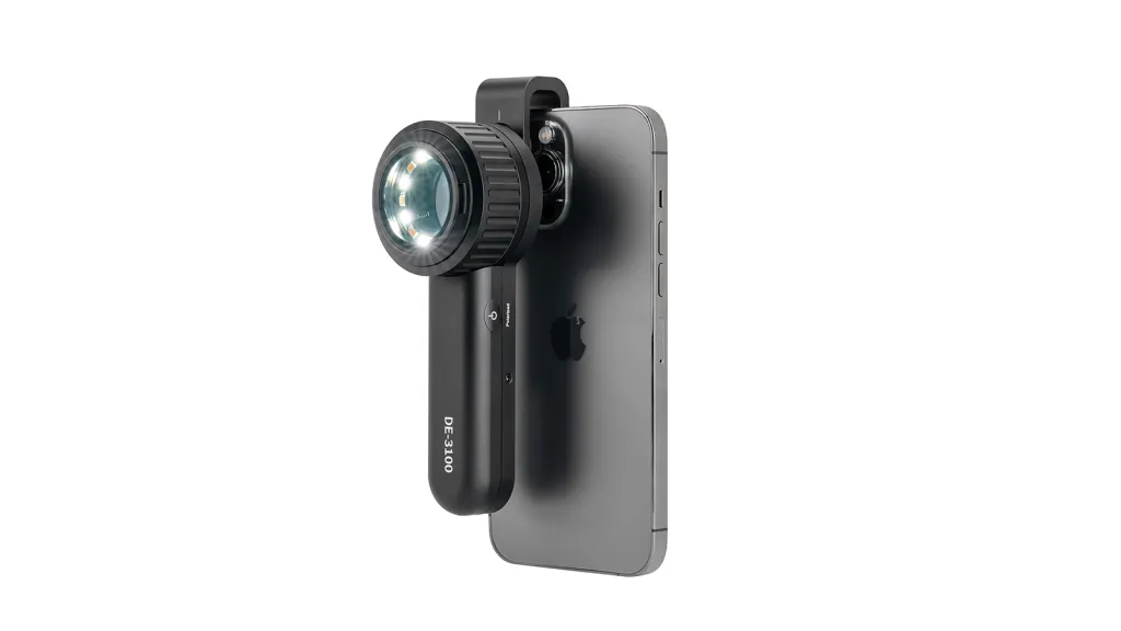
When choosing a dermatoscope, the first thing to consider is the magnification, portability, and lighting system of the dermatoscope, etc. IBOOLO dermatoscopes are made of special glass from Japan, with a light transmittance of more than 98 per cent and uniformity, and both the DE-3100 and DE-4100 have a magnification of more than 10X, which allows for a clear visualisation of the lesions. At the same time, the weight of DE-3100 is only 180g, and that of DE-4100 is only 10X.
DE-4100 weighs only 325g, which is relatively light and easy to carry around for long time use. And both dermatoscopes adopt full metal shell, the appearance of fashionable simplicity, but also better prevent the external bump, to protect the lens body.
Cost-effectiveness is also an important consideration when choosing a dermatoscope, and IBOOLO, as a leading brand of optical dermatoscopes, has been committed to providing the best quality dermatoscopes at an affordable price to its customers. The DE-300 and DE-400 have been well received since their launch.
Dermoscopy in the Diagnosis of Different Skin Diseases
Dermoscopy is important in the diagnosis of many skin diseases. Dermoscopy is useful in the diagnosis of melanoma to some extent by identifying abnormal patterns of pigmentation and vascular distribution. Melanomas show atypical pigment networks, irregular streaks, irregular punctate spherical structures, and irregular vascular patterns on dermoscopy. Blue-white structures, on the other hand, are a relatively specific manifestation of melanoma.
For non-pigmented skin cancers, such as basal cell carcinoma, dermoscopy plays an excellent adjunctive role in detection. Dermoscopy clearly shows brown to bluish greyish-brown radiolucencies, brown to greyish-blue scattered lamellar structures, and multiple bluish-grey globules in basal cell carcinoma. Dermoscopy can also observe the degree of skin inflammation, detect hair diseases, and identify parasitic infections.
IBOOLO Dermatoscope Series Recommendation
The IBOOLO dermatoscope series is divided into pocket dermatoscope series and handheld dermatoscope series. The Pocket Dermatoscope series includes the DE-200, DE-300, and DE-400, which are compact and very easy to carry around. They are relatively simple in function, but they all have polarisation modes and are relatively inexpensive, making them ideal for entry-level learners, or amateurs who want to learn about dermatology on their own.
The IBOOLO handheld dermatoscopes, DE-3100 and DE-4100 series, belong to the high-end optical dermatoscope series. Both models feature a variety of light modes. These include white light polarised, amber white light polarised, amber light, white light unpolarised and the PRO version also has a UV mode. The handheld dermatoscopes are ideal for professional dermatologists, who can use different light modes for different types of lesions. For example, if you want to observe fungal lesions, you can use the UV light mode, where the fungal part of the infection will show a special fluorescent effect.
Tips for Using IBOOLO Dermatoscopes
Before using IBOOLO dermatoscope, firstly, you need to clean the skin surface to be examined, remove dirt, grease and cosmetics, etc., so as not to affect the examination results. Next, gently place the probe of the dermatoscope on the skin surface to be observed, rotate and adjust the focus until the image is clear. Observe the structure and colour changes of the lesions through dermoscopy, and determine the type and degree of progression of the lesions based on the observations.
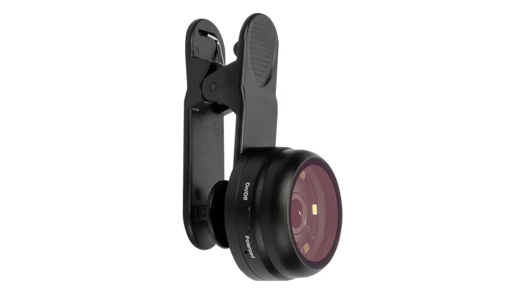
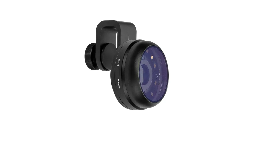
If you want to save the image, you can point the mobile phone clip to the main camera of the mobile phone, then connect the magnetic ring option to the mobile phone clip, and finally connect the dermatoscope magnetically. Switch on the camera function of the mobile phone to save the image.
Immediately after each use of the IBOOLO Dermatoscope, the mirror and handle should be wiped with an alcohol cotton ball or disinfectant wipes to remove any skin flakes, secretions, etc. that may remain. Next, place the dermatoscope in its case for next use.
IBOOLO Dermatoscope Detailed Review
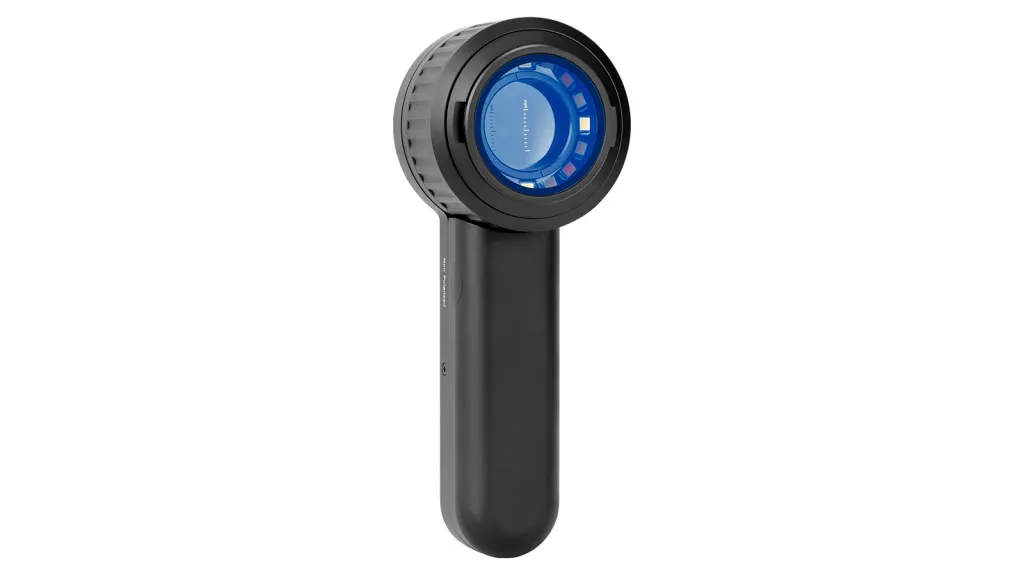
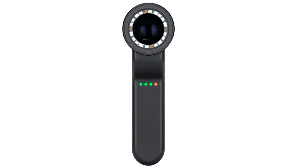
The IBOOLO DE-3100 has a window with a maximum diameter of 32mm and a magnification of 10X and consists of four three-group lenses. Its illumination system consists of 30 LED beads, which can be composed of four different light modes – white polarised, white non-polarised, white amber light and amber light.
The IBOOLO DE-4100 has a window with a maximum diameter of 48mm, a magnification of 10X and consists of four three-group lenses. Its illumination system consists of 22 LED beads, which can form four different light modes – white polarised, white amber polarised, amber and white non-polarised. three brightness adjustments can be made on the DE-4100, allowing the user to select the most appropriate light level according to the brightness of the surrounding environment.
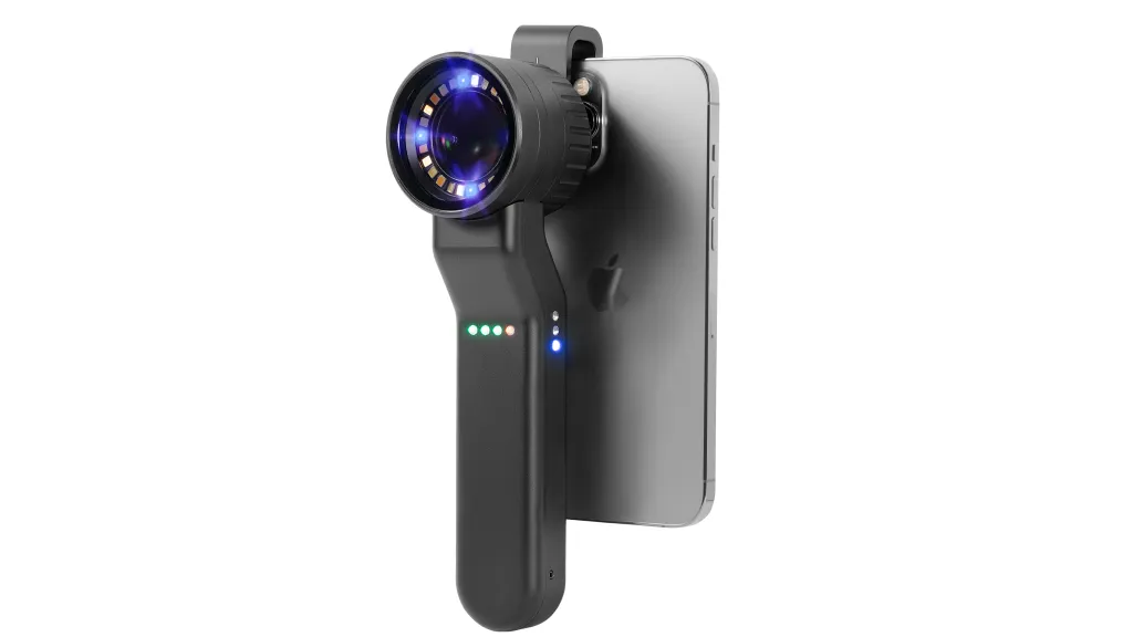
The battery capacity of DE-3100 and DE-4100 is the same 1000 mAh, the DE-3100 can be used for about 7 hours under full charge, and the DE-4100 can be used for about 6 hours under full charge. However, it should be noted that both DE-3100 and DE-4100 can not be charged with fast chargers, otherwise it will cause charging faults, and it is best to use a charger with less than 30W.
The DE-3100 is currently priced at $499 USD and the DE-4100 is priced at $699 USD, product details can be found in the Product section, and any questions are welcome.
Professional Dermatoscope Reviews: A Clinical Guide to IBOOLO Precision Optics
In the field of dermatology, selecting the right diagnostic tool is a decision that impacts both clinical accuracy and patient outcomes. With numerous options on the market, understanding the technical nuances through detailed dermatoscope reviews is essential for modern practitioners. This guide evaluates the IBOOLO product line, focusing on optical clarity, illumination versatility, and ergonomic performance.
The Evolution of Handheld Dermoscopy
Traditional skin examination has moved beyond simple magnification. Modern dermatoscopy requires a sophisticated balance of light manipulation and high-resolution imaging. IBOOLO has positioned itself as a leader in this space by integrating premium Japanese optical glass with advanced LED systems, achieving a light transmittance rate of over 98 percent. This ensures that clinicians can visualize subtle morphological features such as pigment networks, vascular patterns, and crystalline structures with minimal distortion.
Comparative Analysis: IBOOLO Dermatoscope Series
To provide a clear overview for clinicians, we have synthesized technical data into a comparative framework. This allows for an objective look at how different models cater to various clinical needs, from daily screenings to advanced oncological assessments.
| Feature | DE-3100 (Handheld) | DE-4100 PRO (Flagship) | Pocket Series (DE-300/400) |
|---|---|---|---|
| Lens Diameter | 32mm | 48mm (Wide Field) | 25mm - 30mm |
| Magnification | 10x Optical | 10x Optical (Enhanced Depth) | 10x Optical |
| Light Modes | Polarized, Non-Polarized, Amber | Polarized, Non-Polarized, Amber, UV | Polarized / Non-Polarized |
| Weight | 180g | 325g | Under 100g |
| Best For | General Practice | Specialist Oncology | Medical Students / Portability |
Detailed Model Reviews and Clinical Utility
IBOOLO DE-4100 PRO: The Specialist's Choice
The DE-4100 Pro represents the pinnacle of IBOOLO optical engineering. Our technical review finds that its 48mm wide-field lens significantly reduces eye fatigue during long clinic sessions. The standout feature is the inclusion of a UV mode, which is instrumental in diagnosing fungal infections and certain pigmentary disorders that remain invisible under standard white light. The three-level brightness adjustment allows for precise control when examining highly reflective or very dark lesions.
IBOOLO DE-3100: Balance of Power and Ergonomics
Frequently cited in dermatoscope reviews as the most versatile unit, the DE-3100 offers a lightweight 180g frame without compromising on optics. It utilizes a four-lens, three-group system that provides edge-to-edge clarity. For clinicians who prioritize speed, the seamless toggling between polarized and non-polarized modes facilitates the "two-step" diagnostic algorithm efficiently.
The Pocket Series (DE-200, DE-300, DE-400)
For medical students and professionals on the move, the pocket series provides an affordable entry into high-quality dermoscopy. While these units lack the multi-mode complexity of the handheld series, they maintain the essential polarization needed to visualize deep dermal structures and vascularity. Their cost-to-performance ratio makes them a recurring recommendation in introductory dermatoscope reviews.
Technical Considerations: Power and Connectivity
A critical aspect of any professional review is the discussion of real-world usability. Both the DE-3100 and DE-4100 are equipped with 1000mAh batteries, providing 6 to 7 hours of continuous operation. However, a vital technical note for all users: these precision devices must be charged using standard chargers (under 30W). The use of modern USB-C fast chargers can lead to circuit faults, a detail often overlooked in less comprehensive reviews.
Why Digital Integration Matters
In the era of teledermatology, the ability to document and share images is paramount. IBOOLO devices feature universal magnetic adapters that connect to 95% of modern smartphones. This integration allows for high-resolution 4K image capture, enabling clinicians to track lesion progression over time or seek secondary consultations with ease. This digital ecosystem is a major factor in the high satisfaction ratings seen in recent dermatoscope reviews.
Pros and Cons: A Transparent Evaluation
To help you make an informed investment, we have summarized the feedback from our clinical testing panels:
Pros:
- Exceptional optical clarity with minimal chromatic aberration.
- Robust metal construction provides durability in busy clinical environments.
- Highly competitive pricing compared to European and American legacy brands.
- Universal smartphone compatibility enhances documentation workflows.
Cons:
- DE-4100 PRO may feel slightly heavy for clinicians with very small hands.
- Sensitivity to fast-charging cables requires specific charging habits.
Frequently Asked Questions
What should I look for in dermatoscope reviews?
Focus on lens quality, the variety of illumination modes (especially polarization), battery longevity, and how easily the device integrates with digital imaging tools.
Can IBOOLO dermatoscopes be used for trichoscopy?
Yes, the high magnification and polarized light modes of the DE-3100 and DE-4100 are excellent for evaluating hair shaft abnormalities and scalp conditions.
Is the UV mode necessary for all clinicians?
UV mode is highly beneficial for specialists who frequently deal with infectious diseases or specific pigmentary disorders like vitiligo or melasma.
Clinical Disclaimer: This review is based on technical specifications and clinical feedback. Clinicians should choose devices based on their specific diagnostic needs and training.
Recommended reading
High Quality Dermoscopy Meaning Created in Our Products Supply Based in China - IBOOLO
Our China products supply hub couples world-class portability with elite precision, using seasoned expertise to develop high quality dermoscopy meaning for flawless skin visualization anywhere through compact size.
China Skin Cancer Dermoscopy Products Supply Specializes in Professional Items - IBOOLO
Our China products supply creates clinical quality Professional skin cancer dermoscopys enabling powerful skin magnification from anywhere through thoughtful craftsmanship.
China Products Supply Provides Wholesale Dermatoscope Phone Attachments for Clients - IBOOLO
As an expert China products supply, we use exacting wholesale production methods to manufacture high-quality dermatoscope phone attachment solutions tailored for every customer.


