


Article
Dermoscopy Applications in the Diagnosis of Psoriasis
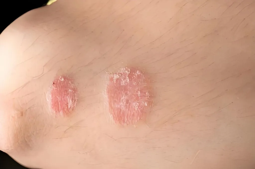
Dermoscopy of Psoriasis What is Psoriasis? Psoriasis is a long term skin disease which can grow anywhere but most commonly appear on the elbows, knees, scalp and trunk. Psoriasis is characterized by rash with itchy, scaly patches. It is a painful chronic disease and with no cure, which means that symptoms appear unexpectedly and may…
Clinical Guide: Advancing the Dermoscopy of Psoriasis Diagnosis
In the rapidly evolving field of dermatology, the dermoscopy of psoriasis has transitioned from a niche research tool to a fundamental bedside diagnostic standard. As chronic inflammatory skin diseases often present with overlapping clinical features, the ability to visualize microscopic morphology is crucial. At IBOOLO, we recognize that precision imaging is the cornerstone of early intervention and long-term management of psoriatic patients.
Psoriasis affects approximately 2-3% of the global population. While clinical diagnosis is often based on the presence of erythematous-squamous plaques, atypical presentations—especially in the scalp, nails, and skin folds—require the superior resolution provided by modern dermoscopy of psoriasis protocols.
The Histopathological Correlation in Dermoscopy of Psoriasis
The hallmark features observed during the dermoscopy of psoriasis are direct reflections of underlying histopathological changes. Understanding this correlation is vital for every practicing dermatologist.
- Regularly Distributed Dotted Vessels: These represent the vertical orientation of dilated and tortuous capillaries within the elongated dermal papillae. In a typical dermoscopy of psoriasis exam, these appear as uniform red dots across the entire lesion.
- White-Silvery Scales: These are a result of parakeratosis (incomplete maturation of keratinocytes) and orthokeratosis. Under magnification, the scales appear thick, lamellar, and highly reflective.
- The Light-Red Background: This hue is the result of significant vasodilation and thinning of the suprapapillary epidermis, allowing the dermal vascular plexus to become more visible.
Clinicians often look for the Dermoscopic Auspitz Sign—where the gentle removal of scales reveals pinpoint hemorrhages. This phenomenon is a definitive diagnostic marker found exclusively through high-quality dermoscopy of psoriasis.
Subtype Variations: Navigating Dermoscopy of Psoriasis Patterns
One of the most complex challenges in clinical dermatology is diagnosing psoriasis variants. The dermoscopy of psoriasis provides unique clues for each:
1. Scalp Psoriasis vs. Seborrheic Dermatitis
Distinguishing between these two conditions is notoriously difficult. However, the dermoscopy of psoriasis on the scalp typically reveals "signet ring" vessels and twisted red loops, whereas seborrheic dermatitis shows more arborizing (branch-like) vessels and greasy, yellowish scales.
2. Nail Psoriasis and the Hyponychium
Nail involvement occurs in up to 50% of patients. Through the dermoscopy of psoriasis in nails, we can observe "pitting" (irregular depressions), splinter hemorrhages, and dilated capillaries at the hyponychium, which are often the earliest signs of psoriatic arthritis.
3. Palmoplantar Psoriasis
Due to the thick stratum corneum on the palms and soles, dotted vessels may be less apparent. The dermoscopy of psoriasis in these areas focuses on white scales following skin furrows and "beaded" vascular patterns.
4. Inverse and Guttate Psoriasis
Inverse psoriasis (found in skin folds) often lacks the characteristic scaling but maintains a bright red background with regular dotted vessels. Guttate psoriasis, often triggered by streptococcal infections, displays a more subtle but still symmetrical vascular distribution during a dermoscopy of psoriasis screening.
Differential Diagnosis: Clinical Accuracy in Dermoscopy
To avoid misdiagnosis and unnecessary biopsies, clinicians must master the differential diagnostic features. The following table highlights why the dermoscopy of psoriasis is the superior diagnostic method.
| Condition | Primary Vascular Pattern | Scale Morphology | Background Color |
|---|---|---|---|
| Psoriasis | Regular Red-Dotted / Glomerular | Thick, Silvery-White | Light Red / Salmon Pink |
| Nummular Eczema | Irregular Dotted Vessels | Yellow Crusts / Serous Exudate | Dull Red / Yellowish |
| Lichen Planus | Wickham Striae / Linear Vessels | Absent or Minimal | Violaceous (Purple) |
| Pityriasis Rosea | Peripheral Dotted Vessels | Collarette (Peripheral) Scaling | Pale Pink |
Optimizing Outcomes with Precision Optics
The effectiveness of the dermoscopy of psoriasis is highly dependent on the quality of the optical lens and lighting. For clinicians, the ability to switch between polarized and non-polarized modes is essential.
Polarization in Psoriasis Diagnosis:Using a polarized dermatoscope like the IBOOLO DE-4100 Pro allows for a deeper view into the dermis without the need for interface fluids. This is particularly useful for visualizing the "dotted vessels" that are the hallmark of dermoscopy of psoriasis.
Furthermore, our smartphone-integrated systems allow for Digital Dermoscopic Monitoring. Clinicians can capture 4K images of psoriatic plaques, providing a quantifiable baseline to measure the efficacy of biologics, phototherapy, or topical steroids. This "dermoscopic healing" often precedes clinical clearance, offering a predictive window for treatment success.
Frequently Asked Questions about Dermoscopy of Psoriasis
Q: Can dermoscopy differentiate psoriasis from mycosis fungoides?
A: Yes. While dermoscopy of psoriasis shows regular dots, mycosis fungoides typically displays fine short linear vessels and a "spermatozoa-like" vascular pattern.
Q: Is dermoscopy necessary for every psoriasis patient?
A: While not always mandatory for clear-cut cases, the dermoscopy of psoriasis is invaluable for atypical presentations, nail involvement, and monitoring therapeutic response to expensive biologics.
Expert Medical Review: This clinical guide on the dermoscopy of psoriasis is curated by the IBOOLO Optical Engineering & Medical Advisory Team.
References:
1. Lallas A, et al. "Dermoscopic patterns of common inflammatory skin diseases." JAAD, 2013.
2. Errichetti E, "Dermoscopy in monitoring the response to therapy in psoriasis." Psoriasis: Targets and Therapy, 2018.
Recommended reading
Top Download DE-3100 User Manual supplier & manufacturer – IBOOLO
IBOOLO is a Top Download DE-3100 User Manual supplier & manufacturer. Information for Download DE-3100 User Manual: DE-3100 user manualDownload...
When will my order arrive – IBOOLO
Most shipments are delivered within 7 days for over 90% of orders on average. All shipping timelines are estimates and while over 90% of orders arrive on time, it does depend on location and customs clearance time frames. Order Processing Times Orders are shipped daily Monday – Friday. Orders placed before the shipping deadline –...
Certificate – IBOOLO
Shenzhen Iboolo Optics Co.Ltd is an experienced manufacturer& exporter in the field of dermatoscope. Our main products include Macro lens, Woods Lamp, Dermatoscope and Microscope. We keep developing varieties of our products and upgrading our quality control systems to enhance our markets competitiveness.
Dermoscopy of Psoriasis
What is Psoriasis?
Psoriasis is a long term skin disease which can grow anywhere but most commonly appear on the elbows, knees, scalp and trunk. Psoriasis is characterized by rash with itchy, scaly patches.
It is a painful chronic disease and with no cure, which means that symptoms appear unexpectedly and may go throughout the whole life. It trends to go through a cycle where it flares for a few weeks or months and then subsides for a while.The condition interferes with sleep and concentration and also varies in severity.
How does dermoscopy detect and diagnose the types of psoriasis skin disease?
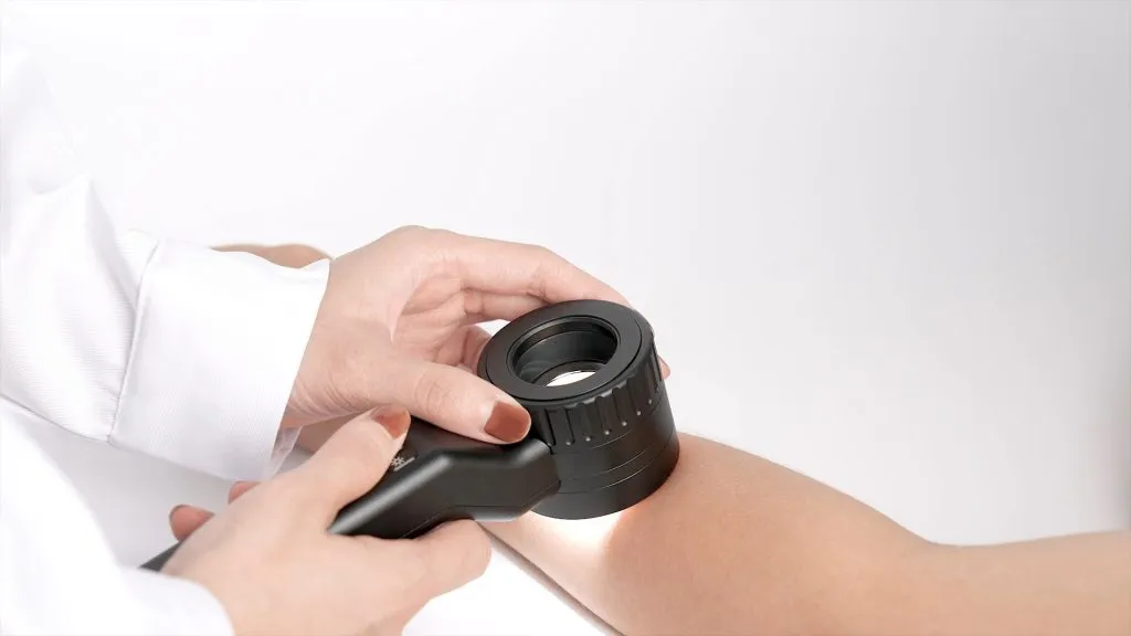
Dermoscopy, also known as dermatoscopy, is a non-invasive tool aiding dermatologists to clearly observe skin lesions which are invisible to the naked eye. To help to diagnose psoriasis, dermoscopy can reveal specific patterns and features of it. The characteristics of dermoscopy of psoriasis as below:
• Dotted vessels: Dotted vessels are the most common dermoscopic features inspected in psoriasis. They show as tiny dots within the psoriatic plaques. If dermscope detect any other morphologic type of vessels, then it can exclude the psoriasis diagnosis.
• Red globules: Sometimes called dots or balls, they correspond to vertically arranged rings of blood vessels within slender dermal papillae. They may differ in diameter, but are usually of similar size within a given lesion.
• Uniform distribution: These vessels at the lesion site showing as symmetrical and uniform distribution is a landmark of psoriatic plaques.
• Removing scales: Removing scales can display tiny red blood drops and reveal the characteristic vascular pattern of psoriasis, called as the dermoscopic” Auspitz” sign.
• Red globular rings: Although red globular rings are rare, but for psoriasis, it is a high specification that circles or rings of red balls present irregularly.
During treatment, dermalogists also can monitor the processing or the transformation of psoriatic plaques with the aid of dermoscope. Thus dermoscopy of psoriasis can provide extra morphological information which may be very useful for early examination of relapse.
How distinguish between psoriasis and eczema under dermoscope?
When using a dermoscope to distinguish between psoriasis and eczema, there are some main characteristics for consideration as below:
Color
Variation: Under dermoscope, psoriatic plaques exhibit a uniform salmon pink color.
Eczema: Eczematous lesions tend to have more various colors, red, yellow, blue or brown are included. These color may change according to the stage of inflammation.
Vascular Patterns:
Psoriasis : Under dermoscopy, psoriasis lesions usually present glomerular blood vessels or regular punctal blood vessel, which is also called “strawberry pattern” ). Most of these vessels are evenly distributed within the lesion part.
Eczema: Eczematous lesions often exhibit more sparse and irregular blood vessels. often showing a linear or linear irregular pattern. These vessels can be less obvious than in psoriasis.
Micro-Hemorrhages:
Psoriasis: Look for pinpoint red dots (micro-hemorrhages) within psoriatic plaques. These dots represent dilated capillaries and are characteristic of psoriasis. Look for tiny red spots (micro-bleeds) in patches of psoriasis. These dots means angiotelectasis, a characteristic of psoriasis.
Eczema: In eczema lesions, micro-hemorrhages are infrequent.
Scale and Crusts:
Psoriasis:Under dermoscope, psoriatic patches usually have silver and white scales with shiny looks.These scales characters thickness and adhesion.
Eczema: Eczematous lesions may appear tiny white scales, and they are less obvious than psoriasis. In addition, eczema lesions may scab due to exudation or scratches.
Distribution and Symmetry:
Psoriasis:Psoriasis patches are usually symmetrically distributed on the surface of the extensor muscles ( knees, elbows, scalp, lower back).
Eczema: Eczema lesions can occur in any part of the body and they are asymmetrical. They may be more common in curved areas (behind the knee,inside the elbow).
There are many key features to distinguish between psoriasis and eczema. Dermoscopy of psoriasis and eczema both can help to enhance the visual field for these skin conditions more closely. So that the dermatologist can make a accurate diagnosis combining dermoscope with clinical experience.
Is dermoscope the main tool for psoriasis diagnosis?
Yes, dermoscope is one of the valuable main tool for psoriasis diagnosis. Dermoscope plays a important role in the process of diagnosing psoriasis. But it is not the sole device for diagnosing psoriasis. There are other devices for helping to diagnose psoriasis more accurate and comprehensive, such as Wood’s Lamp Examination, Laboratory Tests, and Psoriasis Area and Severity Index (PASI).
Other Tools:
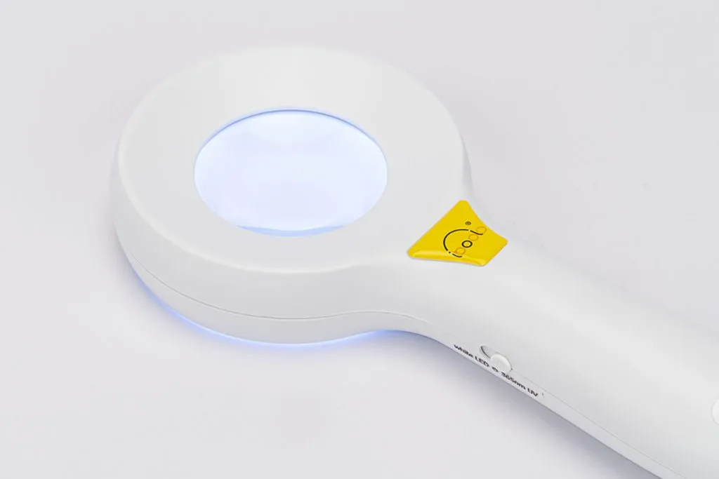
Wood’s Lamp Examination: Wood’s lamp can highlight psoriatic plaques by ultra violet examination because of increased fluorescence.
Laboratory Tests: Blood tests (such as C-reactive protein and erythrocyte sedimentation rate) may provide supporting evidence.Serum markers for autoimmune activity and inflammation are included.
Psoriasis Area and Severity Index (PASI): Based on lesion characteristics, such as the degree of skin severity, the intensity of erythema, scale, and thickness, the PASI assesses the severity of psoriasis.
Additionally, Dermatologists evaluate the patient’s skin by their history, clinical symptoms. Even when it is necessary, a skin biopsy is need to be performed.
While dermoscopy supplies clearly wide visual, a comprehensive approach and other tools are needed to ensures accurate psoriasis diagnosis.
What is the clinical value of dermoscopy in psoriasis?
Dermoscopy a noninvasive device contributing to important clinical value in the evaluation and management of psoriasis. There are some key clinical values for dermoscopy of psoriasis as below:
Psoriasis diagnosis
Identification of Psoriasis: Dermoscopy helps to identify typical features of psoriasis, like regular punctate blood vessels within the erythema plaques.
Timely diagnosis: Dermoscope aids dermologists to diagnose and interfere in timely, and improve the treatment outcome of patients.
Monitoring Disease Progression:
Objective Assessment: Dermoscopy is used to monitor changes in psoriasis over time, and it provides an objective way to assess the severity of psoriasis.
Quantifying Lesions: With using dermoscope, dermatologists can measure the extent of involvement, scaling and erythema of psoriasis.
Treatment Feedback: Dermoscope can monitor precisely the feedback to treatment, even including good fedback, bad feedback and side effect.
Reducing Biopsy Need:
Noninvasive Approach: Dermoscopy greatly reduce the unnecessary biopsies for skin.
Avoiding Invasiveness: Using a dermoscope to examine skin is a invasive and painless process which still can get an accurate diagnosis.
Guiding Treatment Decisions: Targeted Therapies: Under dermoscopy, clinician can choose reasonable treatment therapeutic scheme. Dermoscope is a guider for treatment decisions.
The findings of dermoscopy provide complement information for clinical evaluation. So that clinician can make more proper treatment decisions for psoriasis. Deroscopy of psoriasis is a great significance in examination and management of psoriasis.


