


Article
Dermoscopy of Alopecia Areata
Alopecia areata is a type of hair disease showed hair loss or baldness. It happens when immune system mistakenly attacks hair follicles, causing hair loss. Some hair fall out and then regrow and may fall out again. While some alopecia areata will never regrow hair back. It varies in kinds of complicated situation. Dermoscopy is…
Alopecia Areata Dermoscopy | Precision Diagnosis Unveiled - IBOOLO
Alopecia areata dermoscopy revolutionises hair loss care with precise diagnosis, treatment tracking, and hair restoration guidance. Explore how this non-invasive tool boosts confidence using advanced technology.
Dermoscopy in Alopecia Areata: A Precision Tool for Diagnosis, Monitoring, and Hair Restoration
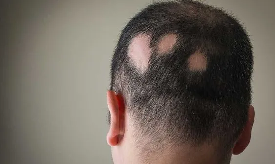
Alopecia areata (AA) is a prevalent autoimmune condition characterised by sudden, non-scarring hair loss in circular or oval patches on the scalp or other hairy areas. In severe cases, it may progress to total scalp hair loss (alopecia totalis) or complete body hair loss (alopecia universalis). Dermoscopy, a non-invasive diagnostic technique, plays a pivotal role in alopecia areata dermoscopy by magnifying scalp and follicular details to reveal hallmark features like “exclamation mark hairs” and yellow dots. This enables clinicians to accurately differentiate AA from conditions such as androgenetic alopecia or scarring alopecia. Beyond diagnosis, alopecia areata dermoscopy is instrumental in monitoring hair regrowth, evaluating treatment efficacy, and guiding hair transplant planning. With advancements in artificial intelligence (AI), dermoscopy’s diagnostic precision continues to improve, offering personalised management for AA patients. This article explores the multifaceted applications of alopecia areata dermoscopy in diagnosis, differential diagnosis, recovery monitoring, and hair restoration, empowering patients to regain confidence.
Understanding Alopecia Areata and Dermoscopy
What is Alopecia Areata?
Alopecia areata is an autoimmune disorder where the immune system mistakenly attacks hair follicles, leading to non-scarring hair loss. Key characteristics include:
- Appearance: Smooth, round, or oval hairless patches on the scalp, with “exclamation mark hairs” often visible at the edges.
- Symptoms: Typically asymptomatic, though some patients report mild itching or burning.
- Risk Factors: Individuals with a personal or family history of autoimmune diseases are at higher risk.
The unpredictable nature of AA, which may resolve spontaneously or progress, underscores the need for precise diagnostic tools like alopecia areata dermoscopy.
What is Dermoscopy for Alopecia Areata?
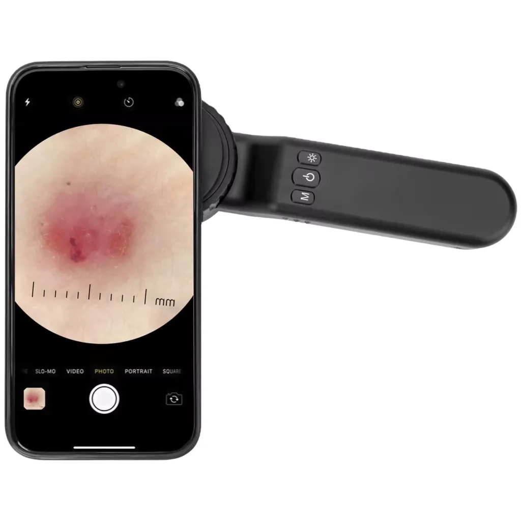
Alopecia areata dermoscopy involves the use of a dermoscope, a handheld device that magnifies scalp and follicular structures to identify specific AA features. By providing a detailed view of hair follicles and scalp patterns, dermoscopy aids in confirming AA, distinguishing it from other hair loss conditions, and assessing disease activity. Its non-invasive nature makes it ideal for repeated use in clinical settings.
Components and Functionality of Dermoscopy in Alopecia Areata
Key Components of a Dermoscope
A dermoscope used in alopecia areata dermoscopy comprises several critical components:
1. Magnification Lens: Offers 10x to 100x magnification to visualise follicular openings, hair shafts, and scalp details.
2. Light Source: Features polarised or non-polarised light; polarised light reduces glare for deeper structural analysis, while non-polarised light highlights surface features.
3. Contact Probe: Ensures stable imaging, with some devices offering non-contact modes for versatility.
4. Imaging Module: Advanced dermoscopes include digital cameras or computer connectivity for image documentation and longitudinal tracking.
5. Accessories: Liquid interfaces (e.g., alcohol or gel) enhance scalp transparency, improving visualisation of follicles and vasculature.
These components work synergistically to enable precise identification of AA-specific features, making alopecia areata dermoscopy a cornerstone of modern dermatology.
How Dermoscopy Works in Alopecia Areata
Alopecia areata dermoscopy operates on principles of optical magnification and optimised lighting:
1. Magnification: High-power lenses reveal micro-features like exclamation mark hairs and yellow dots, critical for AA diagnosis.
2. Lighting: Polarised light minimises scalp reflection, exposing deeper follicular structures, while non-polarised light highlights surface changes like scaling or broken hairs.
3. Imaging: Digital capture allows for consistent monitoring of disease progression and treatment response.
4. Liquid Interface: Applying alcohol or gel reduces surface interference, enhancing the clarity of follicular and vascular patterns.
This technology ensures alopecia areata dermoscopy delivers reliable insights for accurate diagnosis and management.
Standardised Procedure for Alopecia Areata Dermoscopy
The dermoscopy process for alopecia areata is straightforward, painless, and typically performed by dermatologists or hair specialists. The standard workflow includes:
1. Medical History Collection:
- Gather details on hair loss onset, family history, autoimmune conditions, and symptoms like itching.
- Note the number, size, and progression of hairless patches.
2. Visual Inspection:
- Examine the scalp to identify circular bald patches and assess their margins.
- Select key areas for dermoscopic evaluation.
3. Scalp Preparation:
- Clean the scalp to remove debris, oils, or styling products.
- Apply a liquid interface if needed to improve visualisation.
4. Dermoscopic Examination:
- Position the dermoscope close to the scalp, adjusting magnification (typically 10x–50x) and lighting.
- Focus on follicular openings, hair shaft morphology, yellow dots, and exclamation mark hairs.
5. Documentation:
- Capture images for baseline records and future comparisons.
- Analyse findings to assess AA activity or severity.
6. Follow-Up Planning:
Recommend treatments (e.g., topical corticosteroids or immunomodulators) or schedule monitoring visits.
This structured approach ensures alopecia areata dermoscopy is both efficient and reliable.
Applications of Dermoscopy in Alopecia Areata Management
Alopecia areata dermoscopy is versatile, supporting various clinical scenarios:
1. Diagnosis: Confirms AA by identifying exclamation mark hairs, yellow dots, and black dots.
2. Differential Diagnosis: Distinguishes AA from androgenetic alopecia, scarring alopecia, or tinea capitis.
3. Disease Activity Assessment: Evaluates follicular status to determine whether AA is active or stable.
4. Treatment Monitoring: Tracks hair regrowth and scalp changes to gauge therapeutic outcomes.
5. Screening High-Risk Groups: Facilitates early detection in patients with autoimmune or familial predispositions.
6. Research and Education: Provides high-quality images for academic studies and clinician training.
Benefits of Dermoscopy for Alopecia Areata
Alopecia areata dermoscopy offers numerous advantages:
1. High Diagnostic Accuracy: Magnifies subtle features for precise AA identification.
2. Non-Invasive: Painless and safe, ideal for frequent use.
3. Differential Clarity: Differentiates AA from other alopecias, reducing misdiagnosis.
4. Progress Tracking: Assesses disease activity and treatment response over time.
5. Patient-Friendly: Digital records facilitate clear communication and follow-up.
These benefits make alopecia areata dermoscopy indispensable in clinical practice.
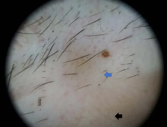
Common Dermoscopic Features of Alopecia Areata
Alopecia areata dermoscopy reveals distinct scalp and follicular patterns:
1. Exclamation Mark Hairs: Tapered hairs, wider at the base and narrower at the tip, indicating active disease.
2. Yellow Dots: Keratin-filled follicular openings, reflecting empty or atrophic follicles.
3. Black Dots: Broken or damaged hair shafts within follicular openings, signalling hair fragility.
4. Short Vellus Hairs: Fine, curly regrowth hairs, marking early recovery.
5. White Dots: Fibrotic or scarred follicles, seen in chronic cases.
6. Scalp Background: Mild erythema in active phases, normalising in stable or recovery stages.
These features, when interpreted alongside clinical history, confirm AA and guide management.
Dermoscopy in Alopecia Areata Recovery Monitoring
During AA recovery, alopecia areata dermoscopy tracks progressive changes:
1. Early Recovery: Increased short vellus hairs and reduced yellow dots, indicating follicular reactivation.
2. Mid-Term Recovery: Thicker, pigmented hair shafts replace exclamation mark hairs, with diminishing erythema.
3. Late Recovery: Restored follicular density and normal scalp texture, resembling healthy skin.
Regular dermoscopic imaging allows clinicians to quantify regrowth and adjust treatments, ensuring optimal outcomes.
Dermoscopy in Hair Transplantation for Alopecia Areata
Alopecia areata dermoscopy enhances hair transplant planning and follow-up, as illustrated in a case study:
Patient Profile: A 35-year-old woman with stable AA patches, seeking transplantation.
Pre-Transplant Dermoscopy:
- Assessed donor site (occipital scalp) for follicular density and hair quality.
- Confirmed disease stability (no exclamation mark hairs or erythema).
- Guided transplant design by mapping recipient areas.
Post-Transplant Monitoring:
- At 1 month: Confirmed graft survival with no inflammation.
- At 6 months: Noted increased hair density and normal shaft diameter.
- Addressed concerns: Yellow dots or atrophy would prompt evaluation for graft failure or AA recurrence.
Alopecia areata dermoscopy thus optimises surgical planning and post-operative care.
Avoiding Misdiagnosis in Alopecia Areata Dermoscopy
Common pitfalls in alopecia areata dermoscopy and their solutions include:
1. Mistaking Normal Follicles for Yellow Dots: Clean the scalp and use polarised light to differentiate keratin plugs from sebaceous material.
2. Confusing AA with Androgenetic Alopecia: Note AA’s focal patches versus AGA’s diffuse thinning, and check for exclamation mark hairs.
3. Overlooking Scarring Alopecia: Identify white dots or follicular dropout, and consider biopsy if scarring is suspected.
4. Misjudging Disease Activity: Combine multiple features (e.g., exclamation mark hairs, erythema) with clinical context for accurate assessment.
Training and adherence to standardised protocols minimise errors in alopecia areata dermoscopy.
Alopecia areata dermoscopy is a transformative tool in the diagnosis and management of AA. By magnifying hallmark features like exclamation mark hairs and yellow dots, it ensures accurate diagnosis and differentiation from other alopecias. Its non-invasive nature supports frequent monitoring of disease activity and treatment response, while its role in hair transplantation enhances surgical precision. With AI-driven advancements, alopecia areata dermoscopy is becoming even more efficient, reducing diagnostic errors and enabling personalised care. From standardised equipment to rigorous protocols, dermoscopy empowers clinicians to deliver evidence-based solutions, helping AA patients restore their hair and confidence.
Alopecia areata is a type of hair disease showed hair loss or baldness. It happens when immune system mistakenly attacks hair follicles, causing hair loss. Some hair fall out and then regrow and may fall out again. While some alopecia areata will never regrow hair back. It varies in kinds of complicated situation. Dermoscopy is a special significant instrument to detect and diagnose alopecia areara.
What is alopecia areata?
Alopecia areata is one of the common types of hair loss. Alopecia areata most typically appearance on the scalp or beard, showing a round or oval bald patch, without hair. And alopecia areata also can happen on anywhere of the body. Alopecia areata is an autoimmune disease which caused by attack from immune system to hair follicles that form hair in skin.
What causes alopecia areate?
When immune system mistakenly attacks hair follicles, leading in alopecia areata. There are not clear reports about what exactly causes the immune attack on hair follicles, but some key factors include:
Genetic Factors: There are genetic factors in the development of alopecia areata due to it has a family tendency.
Autoimmune Factors:The immune system mistakenly targets hair follicles, resulting in hair loss.
Environmental Factors: An exotic virus or another substance happened in the body or other environmental changes may contribute to the development of alopecia areata.
Clinical features of alopecia areate
Alopecia areata usually presents as localized patchy hair loss, ranging from small round patches on the scalp to total body hair loss. It may also affect hair in other parts of the body. The typical clinical features of alopecia areata as following:
Patchy hair loss: Patchy hair loss is the most common form of presentation, with round or oval patches of hair loss and normal skin growth around the patches.
Distribution: Alopecia areata most often occurs on the scalp, but it may also occur in the beard, eyebrows, eyelashes and limbs. Complete follicular openings: The complete follicular openings are directly visible under the dermatoscope.
Positive pull test: Pulling hair will cause hair loss
Terminal hair: In bald patches, terminal hair is completely lost. Others: itching, burning or tingling, no active folliculitis and no symptoms such as erythema, desquamation or pustules around the hair follicles.
Dermatoscopic features of alopecia areata
Dermoscopy is a non-invasive aiding medical instrument that allows precise and tiny details of structures or patterns which are invisible by naked eyes. Dermoscopy of hair, also known as trichoscopy, is ideal and reliable device because it can eliminate light reflection and glare from the surface of the skin. Sometimes it is difficult to distinguish scarring alopecia from non-scarring alopecia, in which case dermoscopy can be really helpful. There are some key dermoscopic features like below:
Yellow spots: Yellow spots can be seen in both acute and chronic alopecia areata and they are yellow circular structures, usually located around the opening of the hair follicle. The presence of yellow spots may be related to inflammation and immune responses.
Black spots: Black spots is a common dermoscopic feature that indicates alopecia areata is in active state . Black spots present broken hairs at the opening of the hair follicle, usually black or brown. They may appear in the center or edges of hair loss patches.
Conical hair: This is a specific dermoscopic feature that shows tiny conical, short and spiky hair tapers gradually toward the scalp, similar to an exclamation point.
Broken hair: Broken hair are short, fractured and broken hairs usually appears in the affected area or the edges of hair loss patches.
Hypopigmentation villi: Hypopigmentation villi are hair that is sparse, short and light in color. This feature usually appears in remission of alopecia areata.
Uses of dermoscope in alopecia areata
Dermoscopy palys a crucial roles in the detection and diagnosis of alopecia areata. There are some typical uses:
1, According to dermatoscopic features of alopecia areata, dermoscopy can accurately distinguish alopecia areata from other types of hair loss, such as scarring alopecias, involutional alopecia, alopecia universalis, trichotillomania, and so on.
2, By dermoscope, skin doctors can exclude other possible causes, like fungal infection, etc.
3, Under the dermoscopy, it is clearly to evaluate the activity and severity of alopecia areata, then to make suitable treatment project
Dermoscopy for the treatment and follow up of alopecia areata
Evaluate the treatment effect: Dermoscopy can give feedback to the treatment effect of alopecia areata, So that skin doctor can analyse and evaluate if the treatment project need to be adjust or not.
Monitor relapse and detect new lesion: Dermoscopy, as a monitor, can inspect the whole process of alopecia areata, including its regrowth, relapse or new lesion.
Cooperate with treatment: By using of dermoscopy and combining with internal and external medication or other treatment methods, it greatly increase the cure effect of alopecia areata.
Notes of the use of dermoscopy in self-examination
It is no doubt that dermoscopy is very useful and reliable device in the diagnosis of alopecia areata. While, there are some notes when people use dermoscopy for self-examination.
Firstly, it is important to rightly operate a dermoscopy and carefully observe the alopecia areata in the process of the examination.
Secondly, skin doctors can compare and analyse the changes of images under dermoscopy, or images photoed by dermoscopy.
Thirdly, when a suspicious finding happened, it is necessary to ask a professional dermatologist for help.
Alopecia areata is a autoimmune skin disease. Even in some cases, hair can regrow back. But it takes undefined time. It may take 6 weeks or 1 year or more long time for hair regrowing. When hair regrow, and it may fall out again. Such situations happen time and again. And some alopecia areata represents, and it will not regrow any more. The development of alopecia areata is very complicated, so it is very important to use dermoscopy to detect and monitor alopecia areata.
No matter patients or skin doctors, all of them should know how to use dermoscopy in right ways. Especially, patients who have alopecia areata diagnosed. Pay more attention to alopecia areata and keep self-examination by dermoscopy. Any suspicious findings, keep in touch with professional skin doctors and cooperate with doctors in diagnosis and treatment.


