


Article
Dermoscopy of Dermal Nevi
Dermal nevi may be present at birth or develop throughout life. These lesions are very common and can present in any individual. Dermal nevi appear in approximately 1% of newborns. Dermoscopy plays an important role as a common observation tool in dermatology that helps doctors observe the deep structure of dermal pigmented nevi. Overview of…
Dermal nevi may be present at birth or develop throughout life. These lesions are very common and can present in any individual. Dermal nevi appear in approximately 1% of newborns. Dermoscopy plays an important role as a common observation tool in dermatology that helps doctors observe the deep structure of dermal pigmented nevi.
Overview of Dermal Nevi
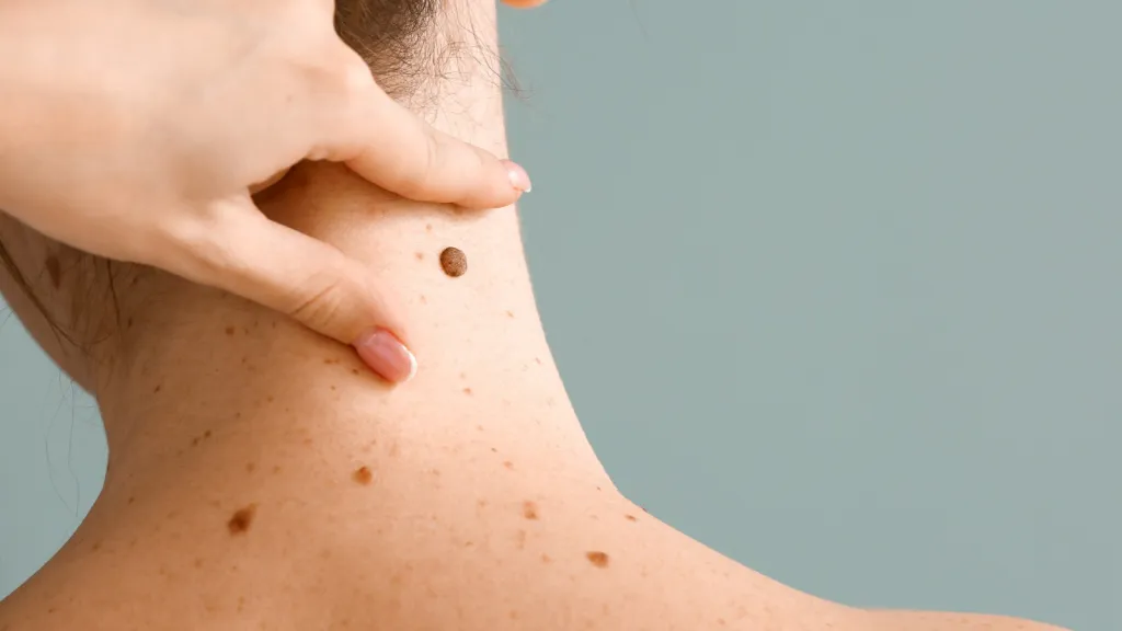
A dermal nevus is a benign, well-defined, raised, colored, papule that appears on the surface of the skin. Dermal nevi can be brown, tan, black, reddish-brown, purple, or skin-colored, and are generally round or ovoid . They may be sessile, raised, and have hair growing from them.
Moreover, dermal nevi are usually benign skin lesions formed by melanocytes in the skin that accumulate in the dermis. Most dermal nevi are stable and do not undergo malignant changes. So, most people don’t need to be alarmed if they find a dermal pigmented nevus on their body.
Principles of Dermoscopy Examination
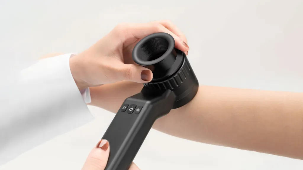
A dermatoscope is a handheld device, equipped with a magnification lens and a light source. It enables the visualization of the subsurface morphology of cutaneous lesions, down to the depth of the superficial dermis. It reveals colors and structures that are normally not visible to the unaided eye and improves the diagnostic accuracy.
Preparation before Digital Dermoscopy Examination
Before the start of dermoscopy, the patient needs to clean the skin surface to be examined in advance, and inform the doctor if there is any local inflammation or breakage, so that the doctor can assess the suitability of dermoscopy. During the examination, the doctor will put the probe of the dermatoscope on the surface of the skin lesion that needs to be observed, keeping a good distance between the probe and the skin. Adjust the light source and magnification of the device until the picture is clear. The doctor will select representative areas of dermal nevus for examination, including the edges and centre of the lesion as well as the surrounding normal skin areas for comparative analysis.
Steps in Dermoscopy
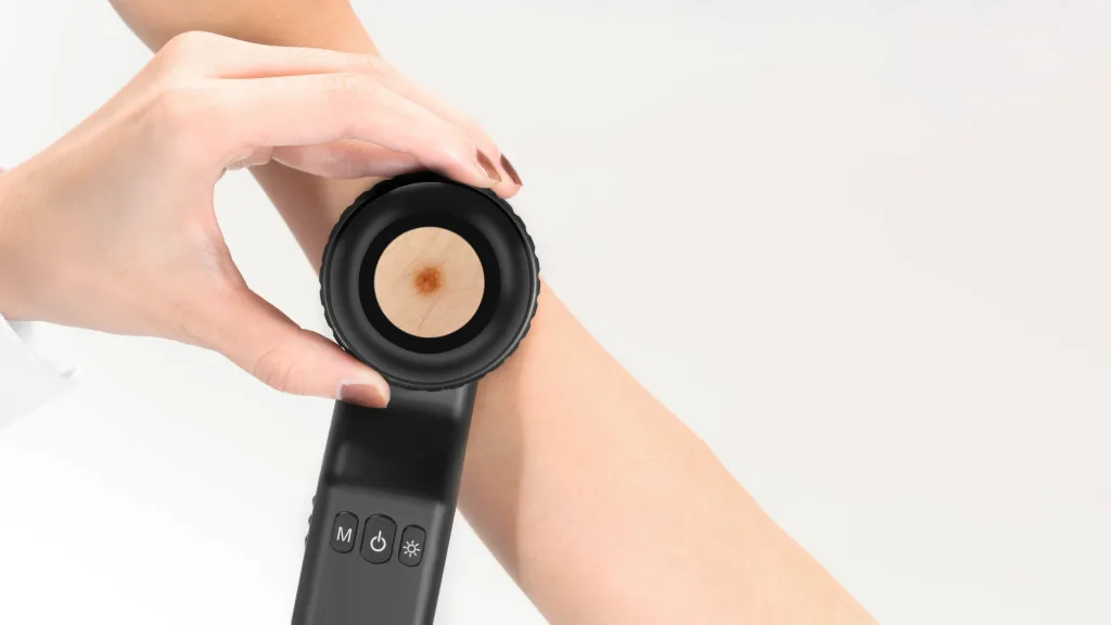
When performing a dermoscopy, first the doctor will select a typical area of dermal nevus skin. Next, gently place the probe of the dermatoscope on the skin surface to be observed, rotate and adjust the focus until the image is clear. Observe the structure and colour changes of the lesions through dermoscopy, and determine the type and degree of progression of the lesions based on the observations. If you want to save the image, you can point the mobile phone clip to the main camera of the mobile phone, then connect the magnetic ring option to the mobile phone clip, and finally connect the dermatoscope magnetically. Switch on the camera function of the mobile phone to save the image. Failure to acquire a dermatoscope image may result if you make a mistake during the operation.
Digital Dermoscopy of Dermal Nevi
The pigment cells of dermal pigmented nevi are mainly distributed in the dermis, usually located in the upper or middle part of the dermis. These pigment cells can be seen dermoscopically usually arranged in clusters, forming islands, curved or scattered within the dermis and even sometimes deep into the subcutaneous tissue. There are several different types of dermal pigmented nevi, including but not limited to:
Flat dermal pigmented nevi:
These nevi are usually smaller, more regular in shape, and commonly found in adult skin.
Nodular dermal nevus:
Nodular dermal naevi are usually larger and often form elevations on the surface of the skin, and can even be felt as hard nodules.
Mixed dermal pigmented nevus:
This type of nevus has mixed features of epidermis and dermis, and pigment cells can be observed under dermoscopy to exist between epidermis and dermis at the same time.
How to Analyse Colours, Patterns, Boundaries and Structures in Digital Dermoscopic Images
Colour:
In dermatology microscopy images, the density and distribution of pigmentation often determines the colour of its appearance. Typically, dermal nevi appear dark brown to black, or lighter if the pigment cells are more dispersed.
Pattern:
Observe the pattern of pigment cell arrangement in the image; normal dermal pigmented nevi appear as regular clusters and may have a more uniform distribution of structures.
Border:
Dermal pigmented nevi usually have clear borders and do not easily penetrate into the surrounding tissues. On the other hand, malignant melanoma, for example, may show irregular, fuzzy borders and may show signs of infiltration or expansion into other layers of the skin.
Structure:
Normal dermal pigmented nevus cells are relatively neatly arranged and the stroma appears normal, whereas abnormal hyperplasia or stromal changes may suggest pathological changes.
Diagnosis and Management of Dermal Nevi
Dermal nevi are benign. Even though benign nevi do not pose health risks such as melanomas, many people opt to have them removed. Most people who seek to have nevi removed do so for cosmetic reasons, for instance, if one is embarrassed about how a particular mole or moles look.
Common methods of removing nevi include: the nevus can be cut off the skin. Some nevi may have subcutaneous cells, which reside underneath the skin, so the doctor might need to make a deeper cut to remove the entire mole to prevent it from growing back. The cut may require stitches.
Differentiation of Dermal Nevi from Other Skin Lesions
Dermal nevi with this clinical morphology will usually reveal one or more of the following dermoscopic features: comma vessels, brown halo, globules, small foci of tan structureless pigmentation, hypopigmented areas. They can also reveal arborizing vessels making it difficult to differentiate them from BCC. The clues to the diagnosis of dermal nevi include the presence of the aforementioned features and lack of other BCC-specific features. In addition, the arborizing vessels in dermal nevi are often a tad out of focus and have a bluish hue. In contrast, in BCC the arborizing vessels are usually sharply in focus and bright red in color.
Application of New Techniques in Digital Dermoscopy
Artificial intelligence (AI) can be defined as the branch of computer science dealing with the simulation of intelligent human behavior in computers. Dermatology has taken the leading position for the implementation of AI in the medical field because of its large clinical, dermoscopical, and dermatopathological image database.
In 2017, Stanford university published a study on deep learning of skin tumors. They trained CNN, using more than 1 lakh images of around 2000 different diseases and tested its performance against that of 21 board-certified dermatologists on biopsy-proven clinical images. It was found that machine had a competence, comparable to that of board-certified dermatologists in identifying and classifying skin cancers.
Conclusion
Dermoscopy contributes to a better visualisation of the deeper components of the skin and assists in the accurate diagnosis of dermal Nevi, including its type, size and distribution, in order to establish more targeted treatment. Doctors can differentiate dermal nevi with other pigmented skin diseases using dermoscopic image analysis and can avoid misdiagnosis after that. Dermoscopy is a promising approach to improving the diagnostic accuracy and efficiency of dermal nevi and other skin diseases with continuous education and technological innovation as important drivers for the continuous development of dermoscopy.
Dermoscopy of Dermal Nevi: A Clinical Guide to Pattern Recognition
Dermal nevi, also known as intradermal nevi, represent the common end-stage of the melanocytic nevus lifecycle. These benign lesions often present as dome-shaped or pedunculated papules, posing a diagnostic challenge when they mimic basal cell carcinoma or other non-pigmented tumors. Utilizing dermal nevi dermoscopy allows clinicians to identify organized micro-architectural patterns that confirm benignity and avoid unnecessary excisions.
Clinical Subtypes and Their Dermoscopic Signatures
In clinical practice, dermal nevi are generally categorized into two main types based on their morphology and location. Recognizing these is the first step in a successful dermoscopy of dermal nevi examination.
1. Unna-Type Nevus
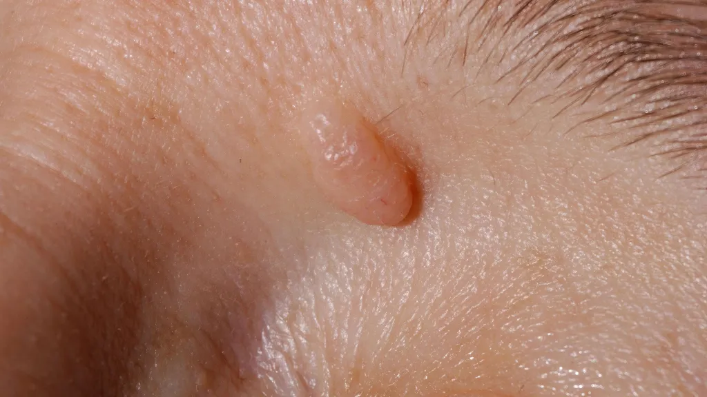
Unna nevi are typically exophytic, soft, and often pedunculated, resembling a raspberry or mulberry. Under the dermascope, these lesions exhibit a "cobblestone" or "globular" pattern. The large, pale-brown globules are clustered together, representing nests of melanocytes safely tucked within the dermis.
2. Miescher-Type Nevus
Miescher nevi are generally firm, dome-shaped, and located on the face or neck. On dermal nevi dermoscopy, these lesions often appear more homogeneous with a pale-tan to skin-colored background. A key feature here is the presence of fine, terminal hairs protruding from the lesion, a strong indicator of a benign nature.
The Diagnostic Hallmarks: Cobblestones and Commas
The reliability of dermoscopy for dermal nevi stems from a combination of specific structural and vascular markers that signify organized growth.
- Cobblestone Pattern: A collection of large, closely aggregated globules that look like a paved road.
- Comma-Shaped Vessels: These are the most common vascular signs in dermal nevi. They appear as small, curved red lines and are usually distributed uniformly throughout the lesion.
- Wobble Sign: When an elevated dermal nevus is gently pushed laterally with the dermatoscope, it shifts easily over the skin surface. This "wobble" suggests a lack of deep invasive tethering.
- Pseudo-network: On the face, pigment may be arranged around hair follicles, creating a mesh-like appearance that should not be confused with the atypical network of melanoma.
Differential Diagnosis: Dermal Nevus vs. Basal Cell Carcinoma (BCC)
Differentiating a non-pigmented dermal nevus from BCC is a frequent clinical requirement. Use the following framework for calibration:
| Feature | Dermal Nevus | Basal Cell Carcinoma (BCC) |
|---|---|---|
| Primary Vessels | Comma-shaped (Uniform). | Arborizing (Tree-like branching). |
| Symmetry | Highly symmetric. | Often asymmetric. |
| Structures | Cobblestones, terminal hairs. | Leaf-like areas, blue-gray nests. |
| Vessel Focus | Often slightly out of focus. | Sharply in focus, bright red. |
Optimizing Analysis with IBOOLO Advanced Optics
To accurately distinguish between comma vessels and the early arborizing vessels of BCC, the clarity of the optical system is non-negotiable. IBOOLO dermatoscopes support the clinician's workflow through:
- 10x Optical Magnification: The industry standard for identifying the "cobblestone" architecture of dermal nevi.
- Cross-Polarization: The IBOOLO DE-4100 Pro allows clinicians to see through the skin surface to visualize the deeper dermal vessels without the need for immersion oil.
- Smartphone Integration: Using the universal magnetic adapter, clinicians can document the lesion's stability over time, ensuring that any subtle growth or vascular change is caught early.
Frequently Asked Questions
Do all dermal nevi have comma vessels?
While extremely common, some very old or non-pigmented nevi may show very few vessels or a completely homogeneous skin-colored pattern.
Can a dermal nevus be removed for cosmetic reasons?
Yes. If dermal nevi dermoscopy confirms the lesion is benign, it can be safely removed via shave excision or laser, often resulting in excellent cosmetic outcomes.
Why is the 'wobble sign' important?
The wobble sign is a simple clinical test that, when combined with dermoscopy, helps confirm that a raised lesion is a superficial dermal nevus rather than an invasive carcinoma.
Recommended reading
High Quality Dermoscopy Meaning Created in Our Products Supply Based in China - IBOOLO
Our China products supply hub couples world-class portability with elite precision, using seasoned expertise to develop high quality dermoscopy meaning for flawless skin visualization anywhere through compact size.
Customized Dermatoscope Iphones Built by Supplier & Manufacturer in China - IBOOLO
With seasoned China-based engineering teams, our supplier & manufacturer company creates specialized dermatoscope iphones to match individual project needs through customized production.
China Skin Cancer Dermoscopy Products Supply Specializes in Professional Items - IBOOLO
Our China products supply creates clinical quality Professional skin cancer dermoscopys enabling powerful skin magnification from anywhere through thoughtful craftsmanship.


