


Article
Dermoscopy of Lentigo Maligna
Lentigo maligna is a form of potentially serious skin cancer, and it is a early stage of lentigo maligna melanoma. Malignant cells of lentigo maligna usually occur in the epidermal layer of the skin. When its malignant cells invade into the dermis or deeper of the skin, then lentigo maligna transforms into lentigo malignant melanoma….
Lentigo maligna is a form of potentially serious skin cancer, and it is a early stage of lentigo maligna melanoma. Malignant cells of lentigo maligna usually occur in the epidermal layer of the skin. When its malignant cells invade into the dermis or deeper of the skin, then lentigo maligna transforms into lentigo malignant melanoma. Therefore, it is particularly important to detect lentigo maligna at its early stage. Dermoscopy is an examination tool that plays an essential role in the diagnosis of early lentigo maligna.
What is advanced lentigo maligna?
Advanced lentigo maligna refers to an advanced stage of lentigo maligna. Lentigo maligna is a early stage of lentigo maligna melanoma. Malignant cells of lentigo maligna usually occur in the epidermal layer of the skin. But advanced lentigo maligna means its malignant cells already have grown from epidermal layer into the dermis or deeper of the skin, it transforms into lentigo malignant melanoma. So advanced lentigo maligna also called as lentigo maligna melanoma.
Clinical features of advanced lentigo maligna (lentigo maligna melanoma)
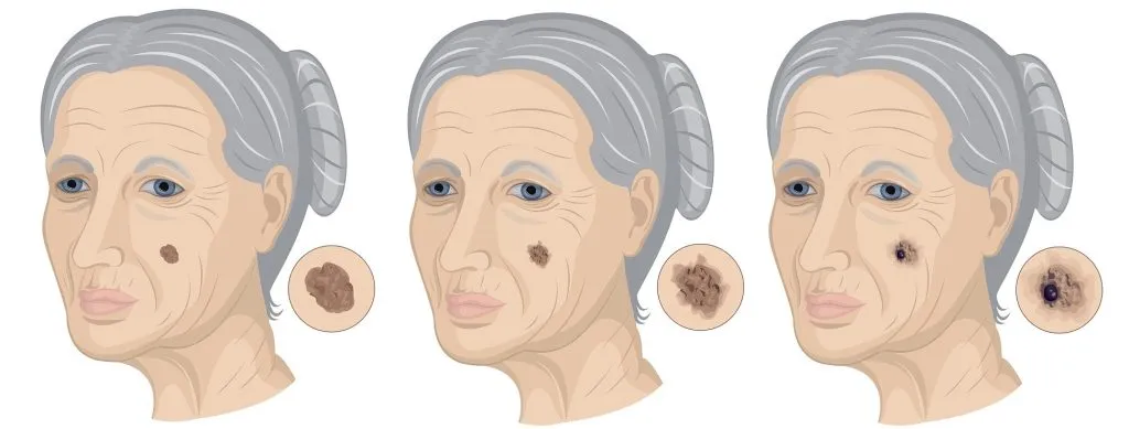
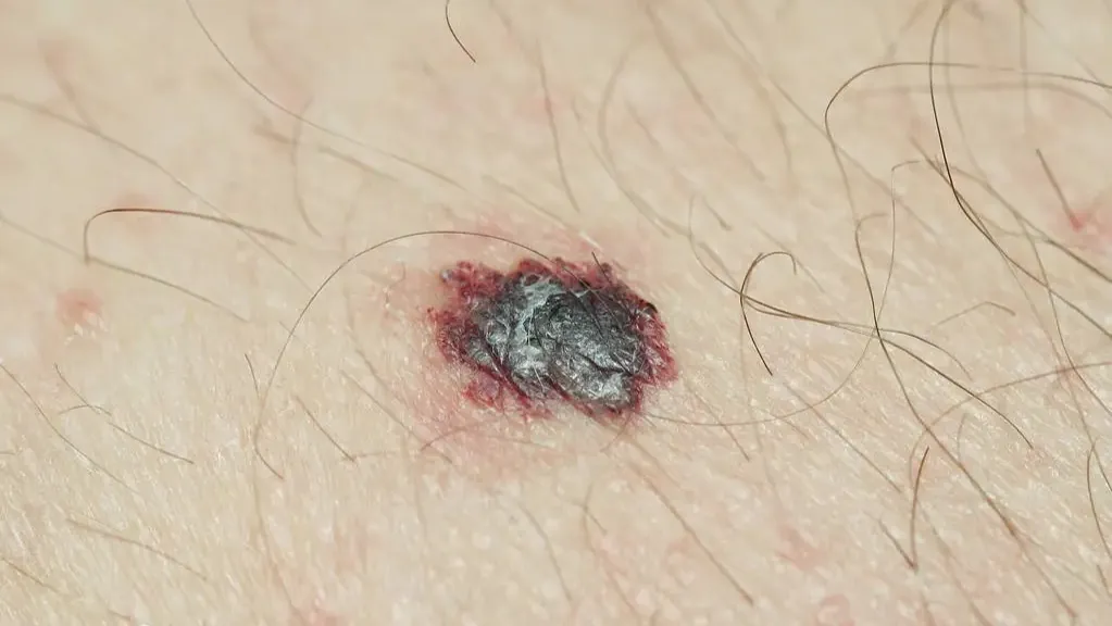
Advanced lentigo meligna usually appears as a large size, irregularly shaped patch of dark skin, varying shades of brown, especially black or blue. It have over thickness of the lesion part, sometimes it may bleed or ulcerate. When it is touched, it shows itching or stinging.It generally develop on sun exposed areas, such as face, neck, nose and arms of middle-age and elder persons, and slightly common on female.
Causes of advanced lentigo maligna (lentigo maligna melanoma)
Sun exposure is a significant high risk factors of advanced lentigo maligna, besides, there are other typical factors cause the diseases of advanced lentigo maligna, like below: Age: As the growing of age, more accumulated sun damage increase the risk of lentigo maligna. That is the reason why lentigo maligna usually happens on middle-age and elder persons. Skin Type: Fair skin people are more likely to have lentigo malgna than dark skin people.
Previous Skin Damage: Previous skin damage will increase the risk of lentigo maligna.
Weakened immune system: People with weakened immune systems have a higher risk of lentigo maligna.
High risk group
Usually, lentigo maligna occurs in middle-age and elder persons more frequently. And the prevalence rate of lentigo maliga of female is slightly higher than male.
Dermatoscopic features of advanced lentigo maligna
As advanced lentigo maligna usually appear as a large, irregularly shaped patch of dark skin in varying shades of brown, black, or blue. Its appearances are similar to other skin lesion, such as seborrheic keratosis, actinic keratosis, etc. Hence it is very difficult to recognize advanced lentigo maligna. At this situation, dermoscopy plays a really crucial roles in the detection and diagnosis of advanced lentigo maligna.
What is Dermoscopy?
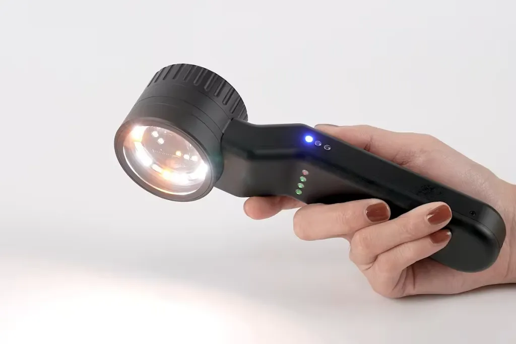
Dermoscopy, also know as dermatoscope, is a handheld aiding device used by dermatologist to observe and diagnose skin lesion and skin diseases, like advanced lentigo maligna. Dermoscopy allows a enhanced visualization field of structures and patters of skin in details by uniting magnifying lenses and powerful lighting system. By using of dermoscopy, dermatologists can make a more accurate and precise analysis and diagnosis of skin lesions.
Dermatoscopic features of lentigo maligna melanoma
There are some certain characteristics of dermoscopy of lentigo maligna melanoma included:
Pseudonetwork on the face: Pigmentation appears around prominent facial hair follicles. Pigmented pseudonetwork is the main features of facial lentigo maligna of dermoscopy.
Asymmetrical pigmented hair follicle openings
Dark rhomboid structure: Proliferation of melanocytes around hair follicle space
Gray spots/lumps/balls: This is the only diagnostic feature in some cases of malignant lentigo)
Dark streaks: lines of melanoma cells in the epidermis or superficial dermis)
Zigzag shape: A series of brown or blue-grey dots forming a zigzag shape.
Others: Color change, irregular and blurry structure and structureless areas are also the main dermoscopic features of lentigo maligna.
Identification of lentigo maligna melanoma by dermoscopy
Senile plaques are typically brown patches and occur on sun exposure areas with multi colors in numbers. But comparison of lentigo maligna, senile plaques are flat, well-defined, and it will not change its size or colors over time. Hence, the dermoscopy takes the role as a monitor to inspect the spots if there is a change or abnormal appearances to identify lentigo maligna from senile plaques.
Due to the looks of lentigo maligna, it brings difficult to distinguish lentigo maglina from other pigmented lesions , such as pigmented actinic(solar) keratosis, seborrheic keratosis, benign lichenoid keratosis by simple naked eye examination. If lentigo maligna is inaccurately diagnosed as a benign skin lesions, like actinic (solar) keratosis which is harmless. It may delay the treatment of lentigo maligna. And let lentigo maligna grow into lentigo maglina melanoma finally. The most useful and noninvasive diagnosis method of lentigo melanoma is to use dermoscopy combining with clinical experience.
The role of dermoscopy in the diagnosis of advanced lentigo maglina (lentigo maligna melanoma)
It is no doubt that a dermoscopy plays a significant role in the detection and diagnosis of lentigo maglina. The key roles lie in such aspects like:
Help skin doctors to make accurate analysis and diagnosis: Under dermoscopy, it allows a enhanced and brightened visual field even some hard-to-reach areas that are invisible by naked eye.
Evaluate the severity of lentigo maglina: Dermoscopy can identify which stage lentigo maglina is in, early stage or advanced stage, then aid skin doctors to evaluate the severity of it.
Monitor the process of lentigo maglina: Dermoscopy is a great help to monitor the growth of lentigo maglina by releasing more clear details of it.
Judge the treatment effect: Dermoscopy can give timely feedback of the treatment effect of lantigo maglina. So that skin doctor can decide if treatment methods need to be adjusted or not.
Precautions for public use of dermoscopy
Dermoscopy is a reliable and helpful technique that helps person to detect and diagnose skin lesions or skin diseases. In professional hands, it is easy to identify a vary of skin diseases. Meanwhile, it is important to use dermoscopy correctly to maximize its effect. There are soem precautions for the use of dermoscopy, the same as the public use of dermoscopy.
Correct operation method of using dermoscopy
How to identify suspicious lesions under a dermoscopy
The need for prompt medical treatment if any atypical lesions are found
Cooperate with doctors for diagnosis and treatment in time
Even though lentigo maglina is a slow-growing cancer. It meas that there is a long way to go from lentigo maglima to lentigo maglima melanoma. But the risk of invasive melanoma is greater when the diameter of lentigo maglina is over 4cm. If left untreated, lentigo maglina may evolve into lentigo maglina melanoma that grows and spreads quickly to other parts of skin. Meanwhile if caught in its early stage of lentigo maglina, there will be a high survival rate of it. So there is really in needed that people pay attention to skin situation and take regular examination. And people should learn and know how to use a dermoscopy correctly for self-examination. Any suspicious findings, have skin checked by professional doctors immediately. Early detection and treatment will bring better health for the skin.
Dermoscopy of Lentigo Maligna | Detection and Diagnosis | IBOOLO
Discover how IBOOLO dermoscopy aids in detecting and diagnosing lentigo maligna. Learn its features, stages, and importance in early skin cancer identification.
Lentigo Maligna Dermoscopy: Early Detection with IBOOLO
Lentigo maligna dermoscopy reveals critical features like annular-granular patterns, enabling early detection of this "hidden killer" skin cancer. IBOOLO’s advanced dermoscopes distinguish it from benign age spots, guiding precise clinical decisions. This article explores key dermoscopic signals, differentiation techniques, and monitoring strategies.
What is Lentigo Maligna?
Lentigo maligna (LM), a subtype of melanoma in situ, often appears as an irregular, brownish patch on sun-exposed areas like the face. Known as a "hidden killer" due to its subtle, age-spot-like presentation, it requires early detection to prevent progression to invasive melanoma. Dermoscopy lentigo maligna provides a non-invasive tool to identify its distinct features.
- Appearance: Flat, unevenly colored patches, often brown or black.
- Common Sites: Face, neck, or other sun-exposed areas.
- Risk Factors: Chronic UV exposure, older age, fair skin.
Role of Dermoscopy in Lentigo Maligna Diagnosis
Lentigo maligna dermoscopy uses high-magnification imaging to reveal microstructural irregularities invisible to the naked eye. IBOOLO’s dermoscopes, with polarised light and digital capture, enhance visualization of dermoscopy lentigo maligna features, ensuring accurate early detection and differentiation from benign lesions.
Three Key Dermoscopic Signals
Dermoscopy lentigo maligna highlights three critical signals for early identification:
- Asymmetric Pigment Network Disruption: Irregular, thickened, or broken network lines, unevenly distributed.
- Gray-Brown Dots/Globules: Scattered, fine granular structures reflecting atypical melanocyte proliferation.
- Annular-Granular Structures: Gray-brown rings around hair follicles, often with central dots forming a target-like pattern.
These signals, observed through lentigo maligna dermoscopy, guide clinicians but require professional interpretation to confirm suspicion of malignancy.
Why These Signals Appear
The asymmetric pigment network in dermoscopy lentigo maligna results from atypical melanocyte growth disrupting the normal grid-like structure. Gray-brown dots/globules reflect scattered melanocyte clusters, while annular-granular structures indicate melanocytes invading along follicular units, a hallmark of LM’s infiltrative behavior.
How Dermoscopy Works for Lentigo Maligna
IBOOLO’s dermoscopy technology employs 10x–100x magnification and polarised light to reduce surface glare, revealing lentigo maligna’s microstructural details. Digital imaging supports documentation for monitoring, making dermoscopy lentigo maligna ideal for non-invasive diagnosis and follow-up.
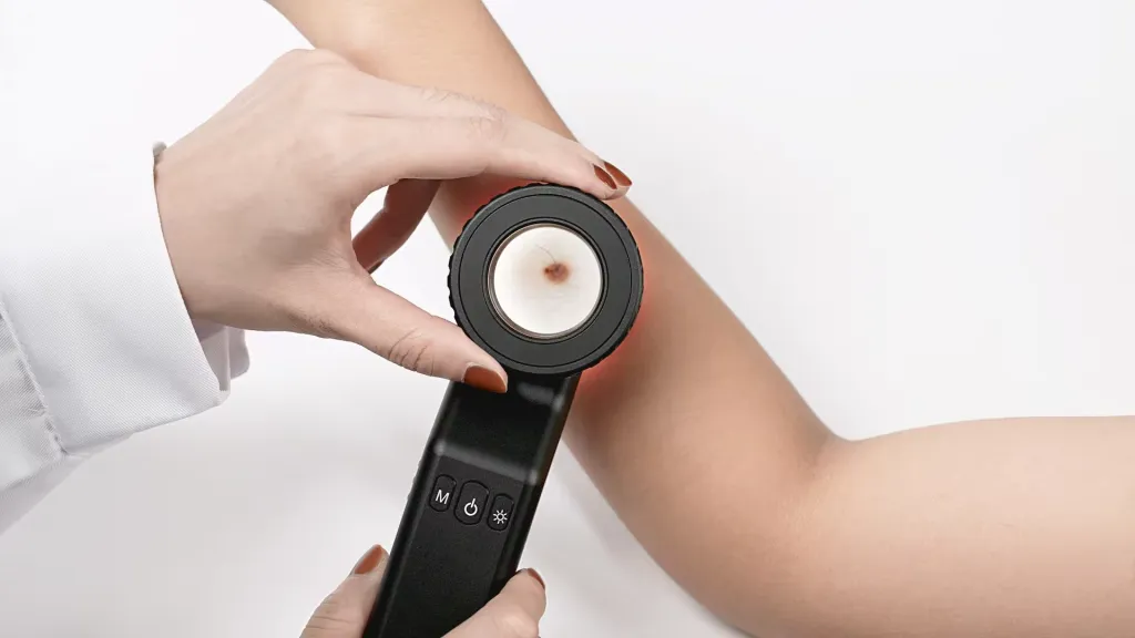
Step-by-Step Dermoscopic Examination
To identify lentigo maligna on dermoscopy, clinicians follow a systematic process:
- History Review: Assess UV exposure, lesion onset, and changes over time.
- Skin Preparation: Clean the area and apply a gel interface for clarity.
- Low-Magnification Scan: Evaluate overall symmetry, color, and border irregularity.
- High-Magnification Focus: Examine pigment network, dots, and follicular structures.
- Image Capture: Record findings for diagnosis and future comparison.
Professional interpretation of dermoscopy lentigo maligna findings is essential for accurate diagnosis.
Differentiating Lentigo Maligna from Benign Age Spots
Lentigo maligna dermoscopy is crucial for distinguishing LM from benign age spots (solar lentigines), guiding appropriate management.
Key Dermoscopic Differences
- Benign Age Spot: Uniform, grid-like pigment network, clear borders, and single-tone color (light brown).
- Lentigo Maligna: Disrupted, asymmetric pigment network, gray-brown dots, annular-granular structures, and jagged, multicolored borders.
Dermoscopy lentigo maligna reveals chaotic, invasive patterns, unlike the orderly structure of age spots. Professional evaluation is critical to avoid misdiagnosis.
Why Self-Diagnosis is Risky
Self-diagnosis using lentigo maligna dermoscopy is unreliable due to overlapping features in atypical cases, the need for high-resolution equipment, and the complexity of interpreting subtle patterns. Only trained dermatologists can integrate dermoscopic findings with clinical history for accurate differentiation.
The Annular-Granular Pattern in Lentigo Maligna
The annular-granular pattern, a hallmark of dermoscopy lentigo maligna, combines gray-brown rings around follicles with scattered granular dots.
Evaluating the Annular-Granular Pattern
- Low-Magnification Overview: Assess overall lesion asymmetry and color variation.
- High-Magnification Focus: Identify gray-brown rings around follicles and scattered dots.
- Pattern Confirmation: Verify the coexistence of rings and granules as a dominant feature.
This pattern, unique to lentigo maligna on dermoscopy, reflects invasive melanocyte growth but requires professional interpretation.
Five Dermoscopic Red Flags for Lentigo Maligna
Lentigo maligna dermoscopy highlights five critical red flags signaling potential malignancy:
- Gray-Brown Dots/Globules: Scattered, irregular granular structures.
- Annular-Granular Structures: Rings around follicles with central dots.
- Asymmetric Pigment Network: Disrupted, uneven network lines.
- Rhomboidal Structures: Polygonal patterns around follicles, often on the forehead.
- Multicolored, Jagged Borders: Mixed colors (brown, gray, black) with irregular edges.
These red flags, observed through dermoscopy lentigo maligna, warrant professional evaluation to confirm malignancy.
Dermoscopy vs. Biopsy for Lentigo Maligna
Lentigo maligna dermoscopy enhances screening but cannot replace biopsy for definitive diagnosis.
Advantages of Dermoscopy
- Non-Invasive: Avoids pain and scarring, ideal for screening.
- Rapid Insights: Reveals microstructural details instantly.
- Monitoring Efficiency: Tracks lesion changes over time.
Dermoscopy lentigo maligna supports early detection and reduces unnecessary biopsies in typical cases.
When Biopsy is Necessary
- Atypical Features: Non-classic patterns or overlap with benign lesions.
- Lesion Changes: Rapid growth, color shifts, or new symptoms.
- Clinical Uncertainty: When dermoscopy cannot rule out malignancy.
Biopsy remains the gold standard, guided by lentigo maligna dermoscopy findings.
Enhancing Lentigo Maligna Care with IBOOLO
IBOOLO’s dermoscopy solutions optimize lentigo maligna management:
- Advanced Dermoscopes: High-resolution imaging with polarised light.
- Clinician Training: Programs to master LM-specific features.
- Patient Education: Informs high-risk groups about screening benefits.
- AI Integration: Enhances accuracy with automated pattern analysis.
Lentigo maligna dermoscopy, powered by IBOOLO’s technology, transforms early detection of this subtle skin cancer. By revealing annular-granular patterns, gray-brown dots, and disrupted networks, it distinguishes LM from benign age spots and supports monitoring. While a vital screening tool, dermoscopy lentigo maligna requires professional interpretation to guide clinical decisions, ensuring optimal patient outcomes.


