


Article
Dermoscopy of Malenoma
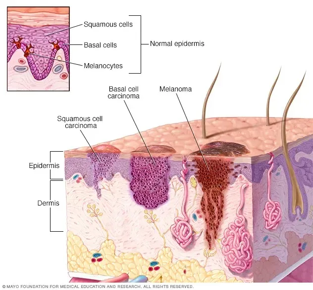
What is malignant melanoma?Malignant melanoma is a serious skin cancer that starts in melanocytes. It is also known as cutaneous melanoma. This skin cancer is much dangerous due to its rapidly spread to other organs if it is not controlled at an early stage.Melanoma can appear in anywhere on the skin, but for people with…
Malignant Melanoma Dermoscopy | Detection Guide - IBOOLO
Expert guide on malignant melanoma dermoscopy by IBOOLO. Discover accurate melanoma detection methods and improve early diagnosis with our dermoscopy techniques.
Melanoma Dermoscopy: A Visual Guide to Early Detection - IBOOLO
Melanoma is the most serious form of skin cancer, but early detection dramatically improves outcomes. This guide explores melanoma dermoscopy, a powerful, non-invasive technique that helps clinicians identify subtle, yet critical, signs of malignancy. Learn how to use this tool effectively and differentiate malignant melanoma dermoscopy features from benign moles.
Mastering Melanoma Dermoscopy: The ABCs of Diagnosis
Dermoscopy acts as a "third eye," revealing subsurface clues that are invisible to the naked eye. When a doctor uses a dermatoscope, they're not just looking at a mole—they're analyzing its intricate micro-architecture. For melanoma dermoscopy, the key is to look for a pattern of disorganization and asymmetry. Here’s a breakdown of the critical features to look for and how to interpret them.
1. Asymmetry in Pattern and Structure
One of the most telling signs of melanoma is a lack of symmetry in its pattern and structure. While a benign mole is often a perfect circle or oval, melanoma is disorganized. Look for:
- Asymmetric Shape: The lesion cannot be neatly divided into two equal halves.
- Asymmetric Borders: The edges are jagged, irregular, and often scalloped or blurred.
- Asymmetric Features: The distribution of colors, vessels, and structures is uneven across the lesion.
This is the first and most critical clue in malignant melanoma dermoscopy.
2. Atypical Pigment Network
The pigment network is the lattice-like pattern created by melanin. In a benign mole, this network is uniform and fades out gradually. In melanoma, it becomes chaotic. Look for a network that is:
- Irregular: The lines vary in thickness and spacing. Some are thick and dark, while others are faint.
- Abrupt Termination: The network stops suddenly at the lesion's edge, rather than fading into the surrounding skin.
- Atypical Coloration: It shows a wide variety of colors, from light brown to dark black, all within the same lesion.
3. Atypical Vascular Patterns
Melanoma requires its own blood supply to grow. Under dermoscopy, these vessels often appear disorganized and abnormal. Key vascular features include:
- Dotted Vessels: Tiny, uniform dots that are chaotically distributed.
- Linear-Irregular Vessels: Serpentine, squiggly vessels that are randomly arranged, a strong sign of malignancy.
- Polymorphous Vessels: A mixture of different vessel types within the same lesion.
These vascular patterns are especially crucial in the diagnosis of amelanotic (non-pigmented) melanomas.
Beyond the Basics: Key Diagnostic Cues
While the ABCs provide a strong framework, melanoma dermoscopy has evolved to include more specific and subtle features that can make a diagnosis more certain.
The Blue-White Veil
This is a hazy, gray-blue or white-blue discoloration within a lesion. It's an ominous sign, as it can represent deep pigment in the dermis or early signs of tumor regression. The presence of a blue-white veil is a strong indicator for biopsy.
Irregular Streaks and Pseudopods
These are irregular, radial projections at the periphery of the lesion. They look like streaks or blunt-ended extensions. Their presence and an asymmetric distribution are highly suspicious and must be biopsied.
Melanoma vs. Benign Moles: A Dermoscopic Comparison
The power of malignant melanoma dermoscopy lies in its ability to differentiate dangerous lesions from harmless ones. Here is a quick comparison table to help you distinguish between the two:
| Feature | Benign Mole (Nevus) | Malignant Melanoma |
|---|---|---|
| Shape & Structure | Symmetrical, organized | Asymmetrical, disorganized |
| Borders | Smooth, well-defined | Irregular, blurred, or scalloped |
| Pigment Network | Uniform, gradually fades out | Irregular, abruptly terminated |
| Vessels | Typically absent or regular | Atypical, dotted, or linear-irregular |
| Colors | One or two uniform colors | Multiple colors (3+), including blue-white |
Why Dermoscopy is a Game-Changer
Studies have shown that dermoscopy can improve melanoma diagnostic accuracy by up to 27% compared to examination with the naked eye alone. This leads to:
- Earlier Detection: Catching melanoma in its earliest, most curable stages.
- Reduced Biopsies: Accurately identifying benign lesions, avoiding unnecessary surgical procedures.
- Enhanced Monitoring: With digital dermoscopy, clinicians can track suspicious moles over time to detect subtle changes.
At IBOOLO, our professional-grade dermatoscopes are built with superior optics and lighting to help you identify these critical features with clarity and confidence. The right technology, combined with a keen eye for patterns, can make all the difference in a patient's outcome.
The Future of Melanoma Diagnosis
The field of malignant melanoma dermoscopy is rapidly evolving. We are now seeing the integration of powerful technologies like Artificial Intelligence (AI) and deep learning algorithms. AI can analyze dermoscopic images to provide a risk assessment score, helping clinicians make faster, more informed decisions. These advancements, combined with regular dermoscopic examinations, are the cornerstone of effective melanoma prevention and management.
Recommended reading
High Quality Dermoscopy Meaning Created in Our Products Supply Based in China - IBOOLO
Our China products supply hub couples world-class portability with elite precision, using seasoned expertise to develop high quality dermoscopy meaning for flawless skin visualization anywhere through compact size.
China Skin Cancer Dermoscopy Products Supply Specializes in Professional Items - IBOOLO
Our China products supply creates clinical quality Professional skin cancer dermoscopys enabling powerful skin magnification from anywhere through thoughtful craftsmanship.
China Products Supply Provides Wholesale Dermatoscope Phone Attachments for Clients - IBOOLO
As an expert China products supply, we use exacting wholesale production methods to manufacture high-quality dermatoscope phone attachment solutions tailored for every customer.
What is malignant melanoma?
Malignant melanoma is a serious skin cancer that starts in melanocytes. It is also known as cutaneous melanoma. This skin cancer is much dangerous due to its rapidly spread to other organs if it is not controlled at an early stage.
Melanoma can appear in anywhere on the skin, but for people with lighter skin, melanoma is more likely to start on the chest and back in men and on the legs in women. The arms, face and neck are other common areas both in men and women. People with darker skin have a lower risk of developing melanoma in these more common areas. Melanoma even can happen in the eyes, while it is rarely to form inside body, like in throat or nose.
What is the cause of melanoma?
The cause of all melanomas is not exactly clear. But melanomas risk increase with exposure to ultraviolet light. Ultraviolet light, also known as UV, comes from sun or solariums. UV radiation can damage the DNA in skin cells, causing mutations that cause the cells to grow uncontrollably and form cancerous tumors.
Melanoma commonly happens especially in tanning, and sunburn during childhood. So it is helpful to reduce the risk of malignant melanoma by limiting the exposure to UV light.
Common high risk groups
The common high risk groups include: a previous melanoma comes back, a history of the sunburns, a family history of melanoma, fair skin or red hair, having many moles or atypical moles, a weakened immune system and so on.
The risk of melanoma appears to be increasing for people under 40, especially women. It is important to understand the high incidence of melanoma in people to help to prevent it. If people find a suspected person, then he should seek medical attention as soon as possible. Melanoma can be successfully treated when it is caught in early stage.
Symptoms of melanoma
There are some common signs and symptoms of melanoma including the following characteristics:
Asymmetry: one half of a mole or birthmark does not match the other half.
Size: Melanomas feature in larger size than normal moles, often exceeding 6mm in diameter.
Color: Melanomas may display different or multiple colors or shades, like brown,black,pink,red,blue, white even mixed color.
Border: melanoma may has irregular, scalloped, notched, or blurred borders.
Pain: Some melanomas may be painful or tender to the touch. Sometimes the spot will bleed and ooze fluid.
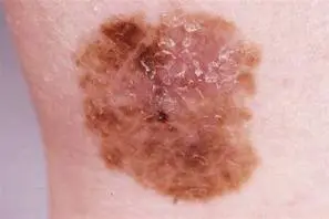
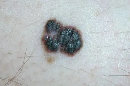
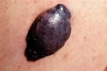
The appearance of new and atypical spots on the skin is the most important warning sign of melanoma. And other important warning sign is the existing spots have changed in color, size and shape. However, there may be one or two unusual characteristics of melanoma. Atypical changes on skin should be pay more attention to.
Survival rates for malignant melanoma
If diagnosed and treated early, malignant melanoma has a high cure rate, and the five-year survival rate for localized melanoma is about 98%. It includes the stage 0, stage I and stage II of malignant melanoma.
If it is not founded in the early stage, and it reaches to stage III of regional melanoma , the survival rate of malignant melanoma is about 63%.
However, if malignant melanoma is not treated until it has reached a more advanced stage and has spread to other parts of the body, such as the liver, lungs or brain is more serious and challenging to treat and has a lower survival rates.The five-year survival rate for stage IV melanoma is about 22%.
No matter how, use a dermoscopy to detect the atypical spots is necessary in case of any chance for evolution of malignant melanoma. Under the dermoscopy, dermatologists can detect and diagnose malignant melanoma much easily and accurately.
Identify the characteristics of malignant melanoma by using dermoscopy
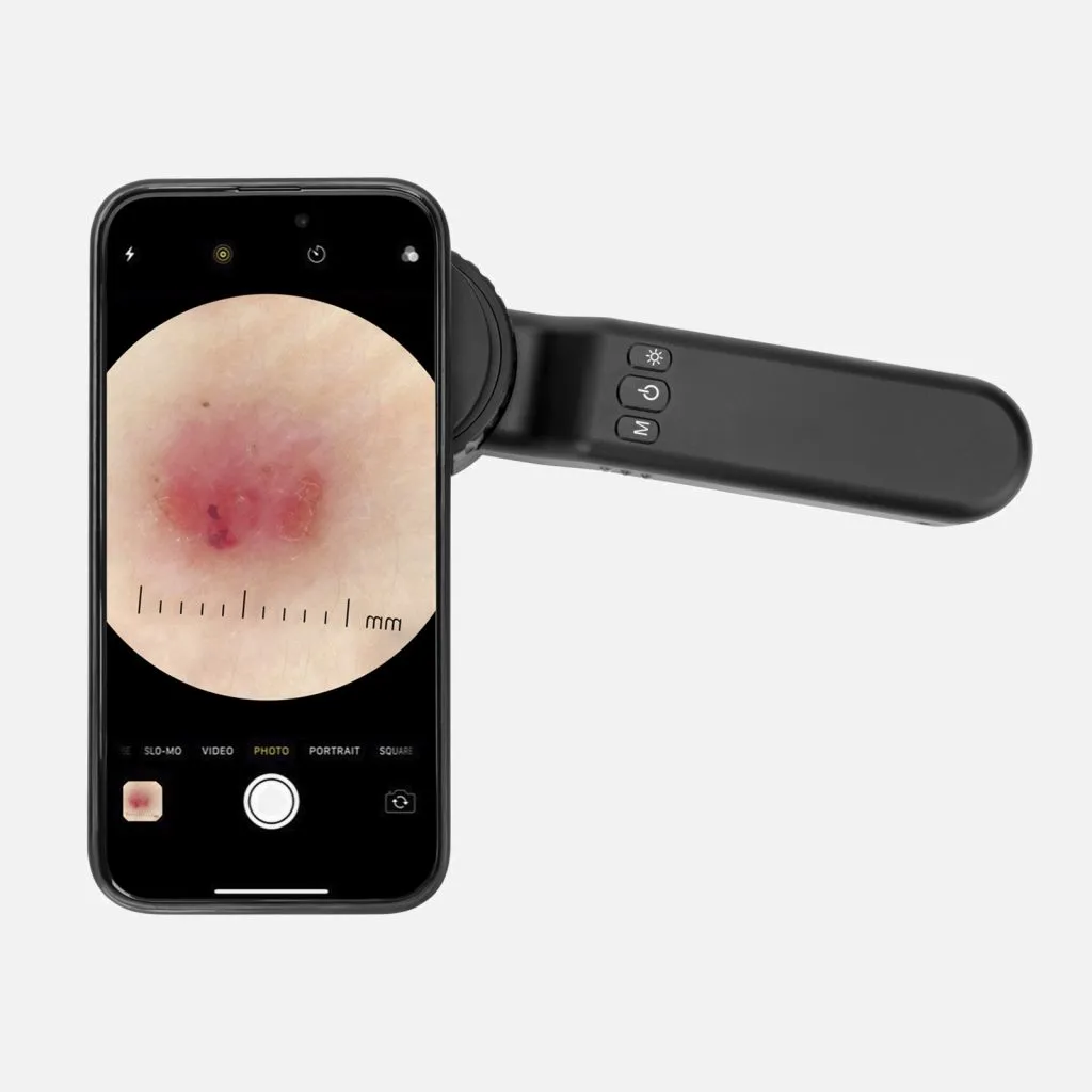
Under the dermoscopy, it is clearly to display the features of malignant melanoma. Such as, by using dermoscopy, malignant melanoma usually clearly shows Irregular edges or borders and multiple colors. And malignant melanoma commonly appears in abnormal pigment distribution and structure, or atypical vascular distribution. Special signs such as granular areas and small blue and white papules may occur. Dermoscopy can precisely distinguish the points of malignant melanoma from benign lesions such as nevus.
Dermoscopy in the diagnosis of malignant melanoma
Dermoscopy is a dependable and aiding medical technique by skin doctors to detect the melanoma. Dermoscopy combines a powerful lighting system and high quality magnify lens to enhance the view of deeper skin and some hard-to-reach locations by naked eyes. The whole process of skin examination under dermoscopy without any side effective or adverse reactions. Dermoscopy can assist professional doctors to make early diagnosis of malignant melanoma without unnecessary biopsies and surgeries. Dermoscopy helps skin doctors to determine the clinical stage of the mass. What’s more, during the whole process of detection or treatment, as a monitor, dermoscopy can observe the progression of lesions. Dermoscopy greatly helps skin doctors to evaluate the effectiveness of surgery or other treatment of malignant melanoma.
Dermoscopy in public self-examination
Dermoscopy is usually designed to a small electric device which is convenient to handheld or easy to take photo by compatible with phones and tablets. Dermoscopy gains a very popularity both in hospital and in home. Dermoscopy can easily identify the malignant melanoma in professional hands. There are some main points when people use dermoscopy in public self-examination.
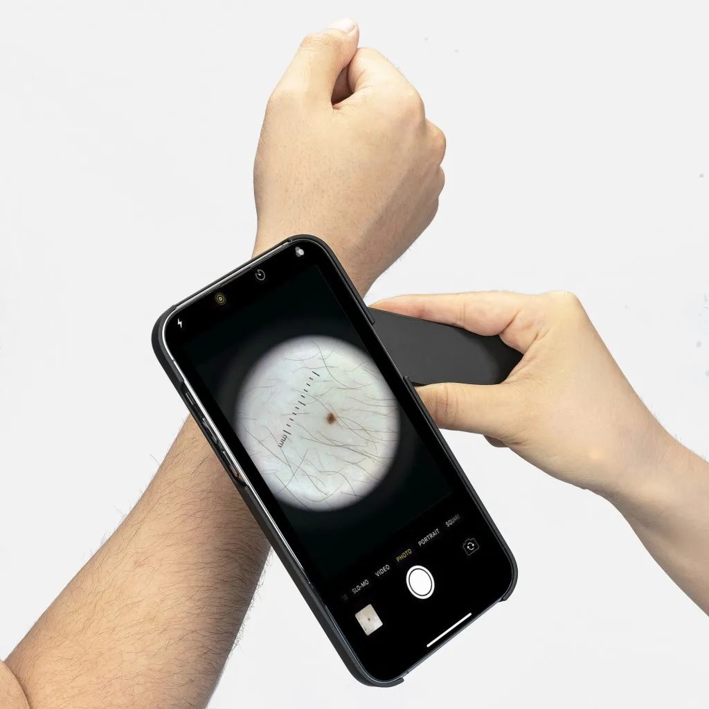
Firstly, correct operation and usage is necessary.
Secondly, during the use of dermoscopy, it needs to keep precautions and observations.
Thirdly, as soon as suspicious lesions are detected by dermoscopy, then it needs to seek medical treatment promptly.
Fourthly, people should cooperate with professional doctors for diagnosis and treatment after self-examination by dermoscopy if any atypical lesions founded.
Skin health is very significant to every one. Detection of malignant melanoma is really important, especially there being negligence in its early stage. With the help of dermoscopy, it increases much more confidence for dermatologists to diagnose and treat malignant melanoma and other skin diseases. In addition, more and more people use dermoscopy to examine skin conditions. Especially, high risk groups of malignant melanoma should even more often use dermoscopy for regular self-examination.


