


Article
Dermoscopy of Molluscum
Molluscum is a worldwide epidemic mainly afflicting children in Papua, New Guinea and Fiji. The molluscum contagiosum virus (MCV) is a DNA virus member of the poxviridae family and is one of human beings’ biggest pathogenic weapons. Dermoscopy, which can magnify and display the lesion morphology in detail (including fine structure such as waxy sheen,…
Molluscum is a worldwide epidemic mainly afflicting children in Papua, New Guinea and Fiji. The molluscum contagiosum virus (MCV) is a DNA virus member of the poxviridae family and is one of human beings’ biggest pathogenic weapons. Dermoscopy, which can magnify and display the lesion morphology in detail (including fine structure such as waxy sheen, peripheral halo or central umbilicus of lesions that suggest Molluscum). These morphologic features become more apparent on dermoscopy and may assist in diagnosis confirmation.
What Is Molluscum?
MCV causes Molluscum, a self-limiting and benign cutaneous infection. Typically, a 2 to 8 mm diameter papule, solitary or multiple; round or hemispherical with waxy luster; central umbilicus concave. Molluscum is transmitted by direct contact, and it may also be spread through autologous inoculation or sexual transmission. Sexual transmission is rampant amongst sexually active youth and often linked with intercourse so that it happens to be considered as a STI.
The disease occurs mainly in children between the ages of 1 and 10 and in people with weakened immune systems, such as those who have leukemia, HIV, or are being treated for cancer. Skin damage can induce viral infection, the virus is easy to enter the human body through the broken skin to become infected. Sharing items with virus carriers in places such as public baths and swimming pools also increases the risk of infection.
How To Identify Molluscum?
Molluscum is caused by a DNA poxvirus called MCV, it does not have an animal host and can only affect humans. It is classified as four types depending on the MCV type, MCV-1 to MCV-4. MCV-1 is the most common type and MCV-2 (found in adults) are typically spread sexually.
Molluscum lesions generally are hemispherical papules, gray or pearly in color, with a waxy surface that has the characteristic central indentation filled with whitish cheese-like grumous material of molluscumor; It is a common skin condition, and because of its symptoms mimic other notable skin conditions such as ordinary warts or papular urticaria or tellest sweat duct tumors.
Numerous combinations of diagnostic approaches for molluscum contagiosum have been developed. We can use dermoscopy to diagnose by a suspected lesion based on typical molluscum contagiosum features first. Histopathological examination can also be used. In the case of a molluscum contagiosum, a centrally depressed dome-shaped cyst filled with keratin is visible beneath the stratum corneum. The presence of molluscum contagiosum can be directly identified and confirmed by gently scraping the cuticle at the tip of the mollusc using a cotton swab and sending the sample to a laboratory to be analyzed for the presence of molluscum contagiosum vesicles.
Dermoscopic Features of Molluscum
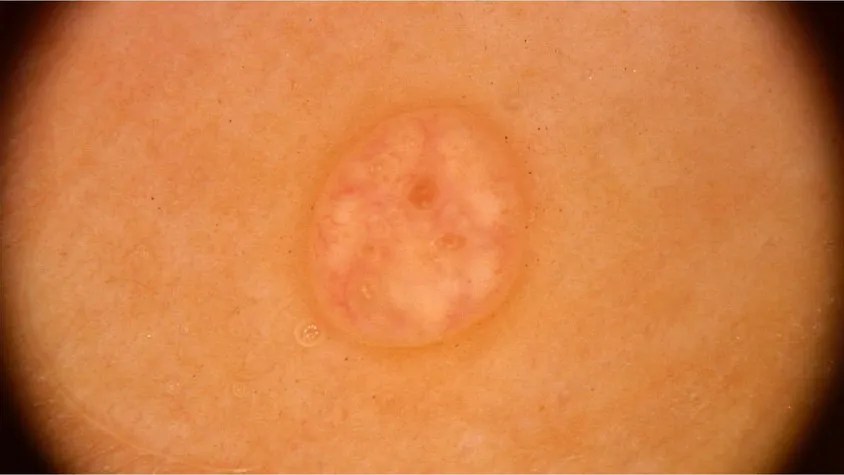
On dermoscopy molluscum shows a central umbilication and the surrounding white to yellow amorphous or multilobulated structures . At the periphery of the lesion, a network of linear or branching blood vessels can be clearly visualized in a distinctive “red crown” pattern, providing a key visual clue to the diagnosis of molluscum contagiosum.
Differences between Molluscum and Other Skin Lesions
Using high-definition magnification with dermoscopy, molluscum contagiosum is easily discriminable from those other lesions of the skin. Central craters and multilobe white to yellowish amorphous structures were common dermoscopic features seen in molluscum contagiosum. The dermoscopic characteristics in basal cell carcinoma include a white structureless background, branching blood vessels and blue-gray dots. Dermoscopic features of keratoacanthoma typically consist of hemispherical or crateriform lesions with a central plug filled by keratin, and often an advancing growth at the margin.
Dermoscopy in the Treatment of Molluscum
Dermoscopy enables real-time observation of changes in molluscum during treatment, including subtle changes in its shape, size and color. For example, molluscum usually appears dermoscopically as an apical depression with a waxy sheen, which may diminish or disappear as treatment progresses, reflecting the effectiveness of the treatment. Furthermore, dermoscopy is a non-invasive procedure that requires no additional pain or discomfort for the patient. This makes it even more popular for monitoring the response to molluscum contagiosum treatment and evaluating efficacy.
Clinical and Dermoscopic Images of Molluscum
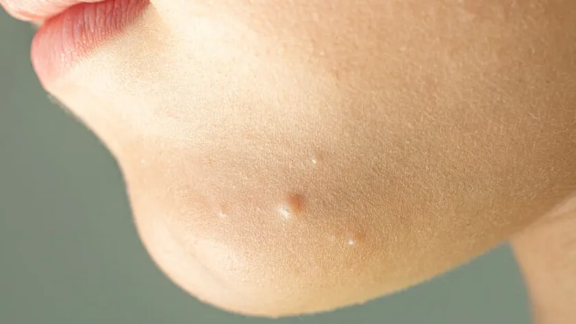
Molluscum contagiosum often presents clinically as a single or multiple translucent papules of varying sizes, mostly skin-colored or slightly whitish, sometimes with an umbilicus at the tip.
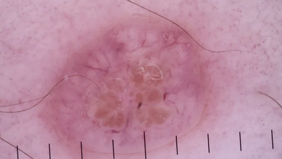
Dermoscopically, molluscum contagiosum lesions often appear as yellowish-white or white amorphous areas, due to the presence of viral particles and keratinized material within the lesions. The lesions are often surrounded by elongated vascular structures, which tend to be crowned or linear, and sometimes punctate.
Dermoscopy clearly shows the fine structure of the lesions and helps the doctor to make an accurate diagnosis. When molluscum contagiosum lesions are atypical or similar to other diseases, dermoscopic images allow doctors to differentiate molluscum contagiosum from other similar diseases (e.g., folliculitis, itchy rashes, sebaceous hyperplasia, etc.), thus avoiding misdiagnosis and underdiagnosis.
Treatment and Management of Molluscum
Common treatments for molluscum contagiosum are divided into two main categories: localized and systemic treatments.
Localized treatments include scraping, freezing and laser treatment. The first step is to use tweezers to scrape or pinch off the warts. The cold treatment is the use of liquid nitrogen’s low temperature effect to make the mollusc tissue necrosis and fall off. Laser treatment uses laser energy to vaporize or carbonize molluscum contagiosum and remove it. The prognosis of all four treatments is also affected by the patient’s immunity, the number and depth of treatments, and post-operative care.
Systemic treatment for molluscum contagiosum focuses primarily on improving the patient’s overall immunity and helping the body to naturally clear the virus. Commonly used immune-modulating drugs for this process include imiquimod cream and recombinant human interferon α2b. Systemic therapy focuses not only on the fading of the lesions, but also on improving the overall health of the patient. By improving the patient’s immunity, systemic therapy can reduce other complications caused by the disease and improve the patient’s quality of life.
Dermoscopy of Molluscum Contagiosum: A Clinical Guide to Identification
Molluscum Contagiosum (MC) is a common viral skin infection caused by a DNA poxvirus. While the diagnosis is often clinical, molluscum dermoscopy has become an essential tool for identifying atypical presentations and distinguishing these lesions from more concerning conditions like basal cell carcinoma or cryptococcosis. This guide explores the hallmark dermoscopic signatures of MC and how advanced optics enhance diagnostic confidence.
The Diagnostic Triad: Recognizing the Poxvirus Signature
The accuracy of molluscum dermoscopy relies on recognizing a specific combination of structural and vascular patterns. When viewed under high-resolution optics, these features reveal the underlying viral colony within the epidermis.
1. Central Umbilication and Pore
The most pathognomonic feature is the central umbilication, which corresponds to the clinical "dimple." Under the dermascope, this indentation often reveals a central pore containing whitish or yellowish amorphous material. This material consists of the "molluscum bodies" (Henderson-Paterson bodies), which are keratinocytes filled with viral particles.
2. Polylobulated White-Yellowish Structures
A key finding in dermoscopy of molluscum is the presence of white-to-yellowish amorphous structures that appear lobulated or "cloud-like." These structures represent the enlarged, infected hair follicle units and are more clearly visualized using the 10x magnification of devices like the IBOOLO DE-4100 Pro.
3. The "Red Crown" Vascular Pattern
The vascular architecture of a molluscum lesion is highly characteristic. Clinicians will typically observe a network of linear, slightly blurred vessels at the periphery of the lesion. These vessels form a ring or "crown" around the central lobules but, importantly, do not cross the center of the lesion—a vital differentiator from malignant tumors.
Differential Diagnosis: Molluscum vs. Mimics
| Feature | Molluscum Contagiosum | Sebaceous Hyperplasia | Common Warts |
|---|---|---|---|
| Central Feature | Central pore with viral bodies. | Central umbilication (duct). | Absent (Papillary surface). |
| Vessels | Red Crown (Peripheral only). | Crown vessels (Thin/Monomorphic). | Dotted/Looped (Thrombus-like). |
| Internal Color | White-Yellow (Opaque). | Creamy-Yellow (Lobular). | Skin-colored or gray. |
Special Considerations: Adult and Immunocompromised Patients
While MC is primarily a pediatric condition, its appearance in adults—particularly in the genital region—requires careful screening for other STIs. In immunocompromised patients (such as those with HIV), lesions may lack the classic central umbilication and appear as large, pearly nodules. Utilizing polarized dermoscopy is critical in these cases to identify faint vascular clues that suggest the underlying viral etiology.
Enhancing Diagnostic Precision with IBOOLO Optics
The subtle "Red Crown" vessels and the texture of internal lobules in molluscum dermoscopy require hardware capable of high-contrast imaging. IBOOLO dermatoscopes support the clinician's workflow through: - High-Resolution Optical Glass: Minimizes chromatic aberration, ensuring the "Red Crown" is accurately colored. - Cross-Polarization: Essential for viewing the deeper vascular patterns without the glare of the waxy lesion surface. - Smartphone Linkage: Allows for teledermatology, enabling pediatricians to share images with dermatology specialists for rapid confirmation.
Frequently Asked Questions
Can dermoscopy distinguish molluscum from Basal Cell Carcinoma (BCC)?
Yes. BCC typically features arborizing (tree-like) vessels that cross the center, whereas molluscum vessels are restricted to the periphery in a crown-like arrangement.
Is it necessary to use immersion oil for molluscum?
While oil can enhance transparency, the cross-polarized light mode on the IBOOLO DE-3100 is generally sufficient and more hygienic for observing viral lesions.
Do all molluscum lesions show a central pore?
Most mature lesions do, but very early or regressing lesions may only show the vascular "Red Crown" or a faint white structureless area.


