Article

Dermoscopy of Nodular Melanoma

Nodular melanoma, as the second most common type of melanoma, makes up around 15 percent to 30 percent of all melanoma cases each year as reported. It is very aggressive due to its faster speed of spread than other types of diseases, especially under the skin where are invisible. High incidence rate, fast growing speed…

Nodular melanoma, as the second most common type of melanoma, makes up around 15 percent to 30 percent of all melanoma cases each year as reported. It is very aggressive due to its faster speed of spread than other types of diseases, especially under the skin where are invisible. High incidence rate, fast growing speed and hidden location bring more difficult to detect and diagnose nodular melanoma. Nodular melanoma causes around 50% of death rate in melanoma-related diseases. But if detected and treated early, there is a great rate to cure it by professional doctors. So it is special crucial to use a dermoscopy to diagnose nodular melanoma in its early stage.

What is nodular melanoma?
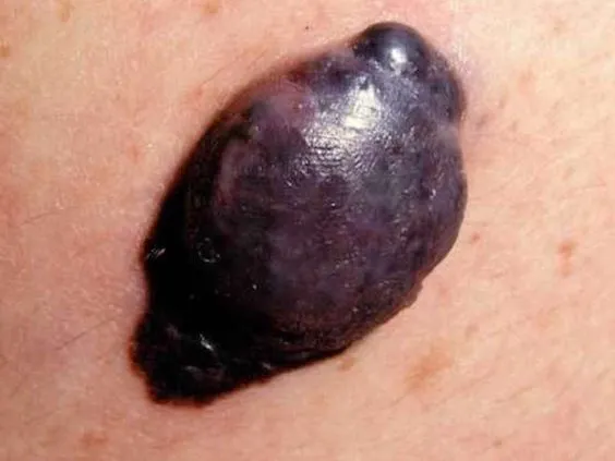
Nodular melanoma is a subtype of melanoma. It usually appears as a firm, discolored bump or node that similar to blood blister above the skin, can be on any part of the body, typically in arms, legs, and trunk.Nodular melanoma is famous as its fast rapid of vertical growing that means it grows quickly in thickness other than in diameter. This is the reason that nodular melanoma recognized as a dangerous and aggressive skin cancer.

What does nodular melanoma look like?
Nodular melanoma usually presents as a round black bump or skin lesion. It looks like a mole, a blood blister, an insect bite, or a skin acne. That’s why people often mistake them for other something other than skin cancer at first. Nodular may also be blue, grey, red, white, pink or brown. Unlike other types of melanomas, nodular melanomas usually have uniform boundaries, symmetrical structure, with the papules rising above the surrounding skin. Symptoms of nodular melanoma usually include itching, tingling, bleeding, and exudation.

nodular melanoma
nodular melanoma

What causes nodular melanoma?
As we all know, liking other factors for melanoma, overexposure to ultraviolet light is also the biggest risk for forming nodular melanoma, especially over sunburns. Except for that, there are several other factors which may cause nodular melanoma, such as: genetic mutation factors, low immune systems, a family history of melanoma, age and gender( the older and man are more risk of nodular melanoma than youth and women), environmental factors, lifestyle factors and so on.

Basic Science of dermoscopy
Dermoscopy is a helpful and valuable device which built in magnification and illumination systems. It allows an enhanced visualization of the surface and subsurface structures of the skin by the basic science of dermoscopy of combining the optical system and physical system. Regarding optical system, dermoscopy usually utilize its cross polarization and non-polarization which present particular dermoscopic features in prominence. Especially it can observe very clear and precise images of structures, patterns and pigment network which are invisible by naked eyes.
Some dermoscopy are designed as a handheld tool which can be hold by skin doctors to observe the skin lesions directly, and also can be used to capture the image for later analysis and comparison. Some advanced dermoscopy are connected with computer software by USB port, detecting and analyzing skin diseases according to dermoscopic features intelligently and effectively.

Dermoscopic features of nodular melanoma
Most melanomas are according to the well-know ABCDE rules of melanoma. But It brings challenge to diagnose nodular melanoma due to its often lacking of these rules. While several typical dermoscopic features of nodular melanoma helping differentiate it from other skin cancers, such as:
Black-blue area or black-blue veil
Atypical vascular patterns
Homogeneous disorganized patterns
Homogeneous blue-black pigmented structure-less areas
Crystalline structures(seen only with polarized light of dermoscopy)
Multiple colors (often more than 3, usually 5~6 colors)
Combination of blue(located in the mid-deep dermis) and black(located in the epidermis)
Combination of polymorphous vessels and milky-red globules/areas
Asymmetrical pigmentation but symmetrical shapes

nodular melanoma dermoscopy

What diseases can be misdiagnosed as nodular melanoma?
Nodular melanoma is characterized as a hard, discolored bump or node that similar to blood blister above the skin. Due to its appearances are similarities with benign and malignant lesions, it challenging to be detected and diagnosed when associated with such skin diseases, like dermatofibroma, hemangioma, pyogenic granuloma, seborrheic keratosis, basal cell carcinoma or angioma.With the help of dermoscopy, it allows doctors to better identify and diagnose the nodular melanoma according to its special dermoscopic features.

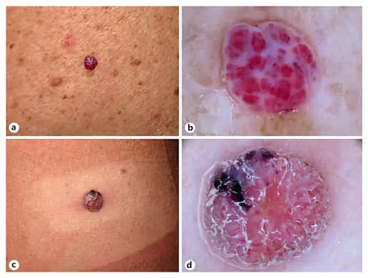
Dermoscopic features of seborrheic keratosis vs nodular melanoma
By using of dermoscopy, it allows more precise and clear visualization of skin lesions. Compared nodular melanoma with seborrheic keratosis, there are some different dermoscopic characteristics between them.
Color:
Seborrheic Keratosis (SK): There is usually white or pale yellow within the seborrheic keratosis.
Nodular Melanoma (NM): There are often 5~6 mixed colors within the lesion,blue-black color is commonly presented.
Shape:
Seborrheic Keratosis (SK):Seborrheic keratosis commonly presents like milary cyst .
Nodular Melanoma (NM): Nodular melanoma commonly presents like blood blister, crystalline.
Structure:
Seborrheic Keratosis (SK): Seborrheic Keratosis with tiny white starry or larger yellowish cloudy.
Nodular Melanoma (NM): Nodular nelanoma with disorganised asymmetrical structure, blue-grey structure, blue-black structure.
Others:
Seborrheic Keratosis (SK): There may irregular crypts,fissures/ridges, blue-grey globules, Light brown fingerprint-like parallel structures, ‘Fat fingers’ be demonstrated in the lesion.
Nodular Melanoma (NM): While, atypical vascular patterns and black-blue veil are the special features within nodular melanoma

the best dermoscopy
the best dermoscopy

Self-examination in nodular melanoma
Since many skin diseases resemble the nodular melanoma, it causes more difficulties to recognize nodular melanoma, especially in its early stage. Hence, it increase the importance to observe and examine by people do self-examination with dermoscopy regularly.
First of all, people should know how to use or operate dermoscopy correctly, and conditions of using dermoscopy, like related working environment. Keeping observing during the process of dermoscopy is also important. If any atypical suspicious findings which are different from normal ones, then the skin conditions should have been checked by professional dermatologists.

Importance of dermoscopy in the diagnosis of nodular melanoma
As the innovative improvement of medical instrument, dermoscopy plays a very important roles in the diagnosis of skin diseases like nodular melanoma. The whole process by dermoscopy to inspect and diagnose skin lesions is noninvasive and painless. Dermoscopy greatly reduces the cross infection during the examinations. And dermoscopy also reduces the unnecessary biopsy and surgery. Dermoscopy becomes a very popular and frequent-used device both among dermatologists and public.


Nodular Melanoma Dermoscopy: Key Features & Diagnosis Guide | IBOOLO

Discover nodular melanoma dermoscopy for early detection of this aggressive cancer. Learn key features like blue-white veil, differentiation from benign nodules, and how IBOOLO’s technology enhances diagnosis.

Nodular Melanoma Dermoscopy: Precision Diagnosis with IBOOLO

Nodular melanoma dermoscopy unveils critical features like the blue-white veil and irregular vascular patterns, enabling early detection of this aggressive melanoma subtype. IBOOLO’s advanced dermoscopes enhance differentiation from benign nodules, guiding clinical decisions. This article explores key dermoscopic signals, high-risk features, and diagnostic strategies for nodular melanoma, emphasizing the need for professional evaluation.

What is Nodular Melanoma?

Nodular melanoma, a highly invasive subtype of melanoma, is characterized by rapid vertical growth, often resembling benign nodules or moles. Its aggressive nature makes early detection critical. Nodular melanoma dermoscopy provides a non-invasive method to reveal microstructural details, aiding timely diagnosis and management.

  • Appearance: Raised, dome-shaped lesions, often black, brown, or skin-toned.
  • Common Sites: Trunk, head, or limbs.
  • Risk Factors: UV exposure, fair skin, family history of melanoma.

Role of Dermoscopy in Nodular Melanoma Diagnosis

Dermoscopy nodular melanoma uses high-magnification imaging to uncover features invisible to the naked eye. IBOOLO’s dermoscopes, with polarized light and digital capture, enhance visualization of nodular melanoma dermoscopy patterns, ensuring accurate differentiation from benign lesions.

Three Critical Dermoscopic Features

Nodular melanoma dermoscopy highlights three hallmark signals for early identification:

  • Blue-White Veil: An irregular, opaque blue-white area, reflecting epidermal hyperplasia and dermal pigment.
  • Irregular Vascular Patterns: Polymorphic vessels (dot-like, linear, hairpin, or tadpole-shaped) with uneven distribution.
  • Asymmetric Pigmentation: Multicolored (black, blue, gray, red) patches with chaotic distribution.

These features, observed through dermoscopy nodular melanoma, guide clinicians but require professional interpretation to confirm malignancy.

Why These Features Occur

The blue-white veil in nodular melanoma dermoscopy results from tumor cell infiltration and epidermal changes, creating an opaque layer. Irregular vessels reflect tumor-induced angiogenesis, while asymmetric pigmentation stems from uncontrolled melanocyte proliferation, hallmarks of malignancy.

How Dermoscopy Works for Nodular Melanoma

IBOOLO’s dermoscopy technology employs 10x–100x magnification and cross-polarized filters to reduce glare, revealing nodular melanoma’s subsurface details. Digital imaging supports documentation, making nodular melanoma dermoscopy ideal for diagnosis and monitoring.

Clinical Workflow for Dermoscopic Evaluation

Clinicians follow a systematic process for nodular melanoma dermoscopy:

  • History Review: Assess lesion onset, growth rate, and risk factors like UV exposure.
  • Skin Preparation: Clean the area and apply a gel interface for clarity.
  • Low-Magnification Scan: Evaluate overall symmetry, color, and border irregularity.
  • High-Magnification Analysis: Focus on blue-white veil, vessels, and pigmentation patterns.
  • Image Capture: Record findings for diagnosis and follow-up.

Professional interpretation of nodular melanoma dermoscopy findings is essential for accurate diagnosis.

The Blue-White Veil in Nodular Melanoma

The blue-white veil, a hallmark of dermoscopy nodular melanoma, appears as an irregular, cloud-like blue-white area, indicating aggressive tumor growth.

Evaluating the Blue-White Veil

  • Low-Magnification Overview: Identify opaque, blue-white areas obscuring underlying structures.
  • High-Magnification Focus: Assess the veil’s uniformity, borders, and extent.
  • Pattern Integration: Confirm its presence alongside irregular vessels or pigmentation.

This feature, unique to nodular melanoma dermoscopy, reflects deep tumor infiltration but requires expert analysis to distinguish from benign mimics like dermatofibromas.

Differentiating Nodular Melanoma from Benign Nodules

Nodular melanoma dermoscopy is crucial for distinguishing malignant nodules from benign lesions like intradermal nevi or basal cell carcinoma, preventing misdiagnosis.

Key Dermoscopic Differences

  • Benign Nodules (e.g., Intradermal Nevi): Uniform pigment networks, comma-shaped vessels, and symmetric structures.
  • Nodular Melanoma: Blue-white veil, polymorphic vessels, multicolored asymmetry, and irregular borders.

Dermoscopy nodular melanoma reveals chaotic, invasive patterns, unlike the orderly features of benign nodules. Professional evaluation is critical.

Five High-Risk Dermoscopic Signals

Nodular melanoma dermoscopy identifies five critical signals warranting immediate attention:

  • Prominent Blue-White Veil: Large, irregular, opaque blue-white areas.
  • Polymorphic Vessels: Mixed vessel types (dot-like, linear, hairpin) with uneven distribution.
  • Multicolored Pigmentation: Three or more colors (black, blue, gray, red, white) in chaotic patterns.
  • Structural Asymmetry: Uneven color and structure across the lesion.
  • Ulceration or Bleeding: Red or black-red areas indicating surface breakdown.

These signals, observed through nodular melanoma dermoscopy, necessitate professional evaluation to confirm malignancy.

Dermoscopy vs. Biopsy for Nodular Melanoma

Nodular melanoma dermoscopy enhances screening but cannot replace biopsy for definitive diagnosis.

Advantages of Dermoscopy

  • Non-Invasive: Avoids pain and scarring, ideal for initial assessment.
  • Rapid Insights: Reveals microstructural details instantly.
  • Monitoring Efficiency: Tracks lesion changes without invasive procedures.

Dermoscopy nodular melanoma supports early detection and reduces unnecessary biopsies in typical cases.

When Biopsy is Necessary

  • Atypical Features: Non-classic patterns or overlap with benign lesions.
  • Rapid Changes: Fast growth, color shifts, or bleeding.
  • Clinical Uncertainty: When dermoscopy cannot rule out malignancy.

Biopsy remains the gold standard, guided by nodular melanoma dermoscopy findings.

Enhancing Nodular Melanoma Care with IBOOLO

IBOOLO’s dermoscopy solutions optimize nodular melanoma management:

  • Advanced Dermoscopes: High-resolution imaging with polarized light.
  • Clinician Training: Programs to master melanoma-specific features.
  • Patient Education: Informs high-risk groups about screening benefits.
  • AI Integration: Enhances accuracy with automated pattern analysis.

Nodular melanoma dermoscopy, powered by IBOOLO’s technology, transforms the diagnosis of this aggressive melanoma subtype. By revealing blue-white veils, polymorphic vessels, and asymmetric pigmentation, it distinguishes nodular melanoma from benign nodules and guides clinical decisions. While a vital screening tool, dermoscopy nodular melanoma requires professional interpretation to ensure accurate diagnosis and optimal patient outcomes.

Share this article

0

No products in the cart.

Ask

Have questions on gear or your order?

Our Gear Guides are here to help! Get personal advice from pro creatives

Name
Subject
Email address
How can we help?
Answers

Instant Answers

How can dermoscopy images be captured?

Dermoscopy images can be captured and stored in different ways, such as: • Using a smartphone or tablet with dermoscopic adapter, which consisted in the package.• Using a digital camera

Dermoscopy images can be captured and stored in different ways, such as:

• Using a smartphone or tablet with dermoscopic adapter, which consisted in the package.
• Using a digital camera with dermoscopic adapter, there’s 49mm screw size camera adapter available to order now.

Compatible phone/tablet models:
All iPhone models, 95% Android phones, 90% tablet. For phone/tablet size in 5.25-14mm

Compatible camera models:
All camera with built 49mm filter screw, such as Canon EOS 70D, 80D, 90D; Canon EOS R7, R10, R50, R100; Canon M100, M200, M50, Mark II; Canon G7X Mark III, Sony ZV-1

How can I connect my phone to my dermatoscope?

There’s universal phone adapter for all our dermoscopes. Please check the installation procedure bellow or watch operation guide. Smartphone Connector (1) Place phone adapter screw in the center of smartphone’s

There’s universal phone adapter for all our dermoscopes. Please check the installation procedure bellow or watch operation guide.

Smartphone Connector

(1) Place phone adapter screw in the center of smartphone’s main camera.
(2) Screw magnet attachment on phone adapter.
(3) Put dermoscope’s back ring and magnet attachment together

Take The Best Images

You need to adjust the focus ring after the dermoscpe connected on smartphone to get the best images.

How can I clean my dermoscopy after usage?

Cleaning your dermoscopy after usage is important to prevent cross-contamination and infection. The cleaning method may vary depending on the type and model of your dermoscopy, so you should always

Cleaning your dermoscopy after usage is important to prevent cross-contamination and infection. The cleaning method may vary depending on the type and model of your dermoscopy, so you should always follow the manufacturer’s instructions. However, some general steps are:

• Turn off and disconnect your dermoscopy from any power source or device.

• Wipe off any visible dirt or debris from the dermoscopy with a soft cloth or tissue.

• Disinfect the dermoscopy with an alcohol-based wipe or spray, or a disinfectant solution recommended by the manufacturer. Make sure to cover all surfaces, especially the lens and contact plate.

• Let the dermoscopy air dry completely before storing it in a clean and dry place.

• Do not use abrasive or corrosive cleaners, solvents, or detergents that may damage the dermoscopy.

• Do not immerse the dermoscopy in water or any liquid, unless it is waterproof and designed for immersion.

You should clean your dermoscopy after each use, or at least once a day if you use it frequently. You should also check your dermoscopy regularly for any signs of damage or malfunction, and contact the manufacturer or service provider if needed.

Polarized VS Non-polarized Dermoscopy

A dermoscopy is a device that allows the examination of skin lesions with magnificationand illumination. By revealing subsurface structures and patterns that are not visible tothe naked eye. It can

A dermoscopy is a device that allows the examination of skin lesions with magnificationand illumination. By revealing subsurface structures and patterns that are not visible tothe naked eye. It can improve the diagnose accuracy of skin lesions, such as melanoma,basal cell carcinoma, seborrheic keratosis, etc.

There are two main types of dermoscopy: Non polarized and polarized dermoscopy.We’ve fitted most of our dermoscopys with polarized and non-polarized light. They canbe used in multiple skin structures.

Non-polarized contact Mode

In non-polarized mode, the instrument can provide information about the superficialskin structures, such as milia-like cysts, comedo-like openings, and pigment in theepidemis.

The dermoscopy requires applying a liquid such as mineral oil or alcohol to the skin andplacing the lens in contact with the skin. This reduces surface reflection and enhancesthe view of subsurface structures.

Image with non-polarized light (DE-3100)

Polarized contact Mode

In polarized mode, the instrument allows for visualization for deeper skin structures,such as blood vessels, collagen, and pigment in the dermis.

The dermoscopy does not need to be in contact with the skin or use any liquid. Theirpolarized light can help to eliminate surface reflection and allow visualization ofvascular structures.

Image with polarized light (DE-3100)

Polarized non-contact Mode

The dermoscopy can also use polarized light to examine the skin without direct contact.

In polarized non-contact mode, the instrument allows for examination infected areasand lesions that are painful for the patient, or the difficult to contact pigmented lesions,such as nails and narrow areas.

The contact plate should be removed in this mode, and it does not require applying aliquid to the skin. As it doesn’t require pressure or fluid application on the skin, it canalso avoid cross-contamination and infection risk.

Image in polarized non-contact mode (DE-3100)

How effectiveness is dermoscopy

Compared with visual inspection, the dermoscopy can be used to capture and store skin lesion photos, which play an important role in early skin cancer examination. The dermoscopy allows the

Compared with visual inspection, the dermoscopy can be used to capture and store skin lesion photos, which play an important role in early skin cancer examination.

The dermoscopy allows the examination of skin lesions with magnification and illumination. This can be greatly avoiding the factors that cause interference to visual detection. Such as lighting, skin color, hair and cosmetics.

Several studies have demonstrated that dermoscopy is useful in the identification of melanoma, when used by a trained professional.

It may improve the accuracy of clinical diagnosis by up to 35%
It may reduce the number of harmless lesions that are removed
In primary care, it may increase the referral of more worrisome lesions and reduce the referral of more trivial ones

A 2018 Cochrane meta-analysis published the accuracy of dermoscopy in the detection.

Table 1. Accuracy of dermoscopy in the detection of melanoma in adults
Detection Method Sensitivity, % Specificity, % Positive Likelihood Ratio NegativeLikelihood Ratio
Visual inspection alone (in person) 76 75 3.04 0.32
Dermoscopy with visual inspection (in person) 92 95 18 0.08
Image-based visual inspection alone (not in person) 47 42 0.81 1.3
Dermoscopy with image-based visual inspection (not in person) 81 82 4.5 0.23
ROC—receiver operating characteristic. *Estimated sensitivity calculated on the summary ROC curve at a fixed specificity of 80%.

As we can see, the dermoscope can improve the accuracy of diagnosis of skin lesions, especially melanoma.

Table 1. Accuracy of dermoscopy in the detection of melanoma in adults
Detection Method Sensitivity, % Specificity, % Positive Likelihood Ratio NegativeLikelihood Ratio
Visual inspection alone (in person) 79 77 3.4 0.27
Dermoscopy with visual inspection (in person) 93 99 93 0.07
Image-based visual inspection alone (not in person) 85 87 6.5 0.17
Dermoscopy with image-based visual inspection (not in person) 93 96 23 0.07
ROC—receiver operating characteristic. *Estimated sensitivity calculated on the summary ROC curve at a fixed specificity of 80%.

Characteristics of the dermatoscopic structure of the skin lesions include:

• Symmetry or asymmetry
• Homogeny/uniformity (sameness) or heterogeny (structural differences across the lesion)
• Distribution of pigment: brown lines, dots, clods and structureless areas
• Skin surface keratin: small white cysts, crypts, fissures
• Vascular morphology and pattern: regular or irregular
• Border of the lesion: fading, sharply cut off or radial streaks
• Presence of ulceration

There are specific dermoscopic patterns that aid in the diagnosis of the following pigmented skin lesions:

• Melanoma
• Moles (benign melanocytic naevus)
• Freckles (lentigos)
• Atypical naevi
• Blue naevi
• Seborrhoeic keratosis
• Pigmented basal cell carcinoma
• Haemangioma

We use cookies on this website to provide a better user experience. By continuing to browse the website, you are giving your consent to receive cookies on this site. For more details please read our Privacy Policy.

Hot Search Terms