


Article
Dermoscopy of Scalp Psoriasis
Scalp psoriasis, a common chronic, recurrent autoimmune disease, has a definite genetic predisposition to scalp psoriasis. Its prevalence is reflected in its wide spectrum and high incidence. The scalp, due to its special physiological position, is poorly insulated and susceptible to wind-cold, thus increasing the probability of developing the disease.Dermatoscope plays an important role in…
Scalp psoriasis, a common chronic, recurrent autoimmune disease, has a definite genetic predisposition to scalp psoriasis. Its prevalence is reflected in its wide spectrum and high incidence. The scalp, due to its special physiological position, is poorly insulated and susceptible to wind-cold, thus increasing the probability of developing the disease.
Dermatoscope plays an important role in the diagnosis and treatment of scalp psoriasis. Dermoscopy is able to magnify fine skin structures. Through dermoscopy, doctors are able to observe subtle changes on the surface of the scalp, including psoriasis, intense erythematous reactions, follicular orifice plugs and burrs.
Overview of Scalp Psoriasis
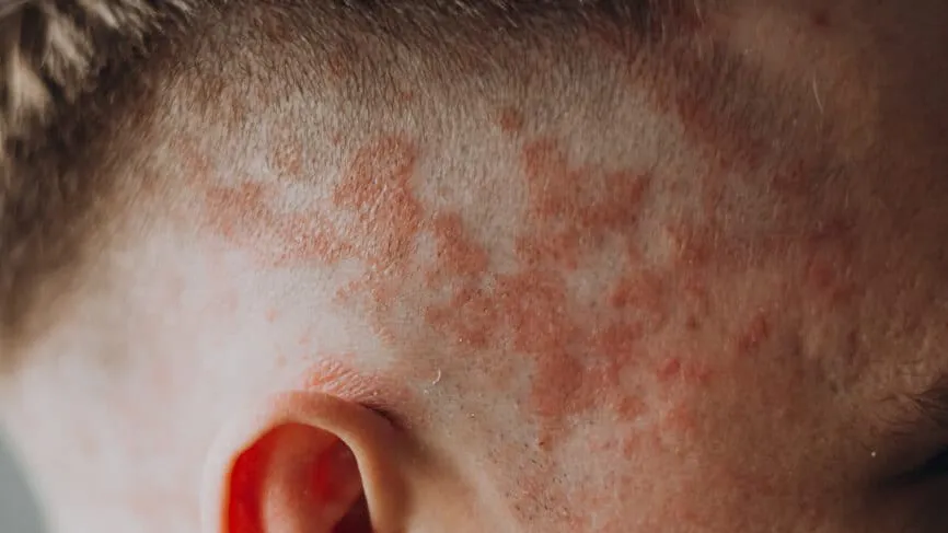
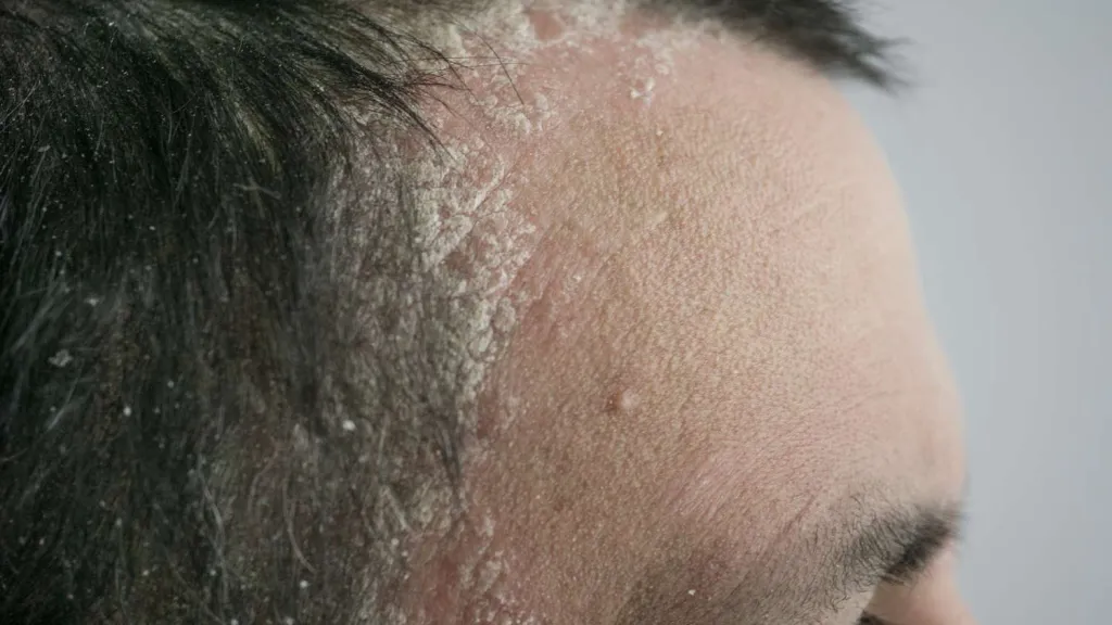
Scalp psoriasis is a common chronic inflammatory skin disease characterised mainly by the appearance of reddish patches on the scalp and these patches are covered with silvery-white scales and accompanied by symptoms such as itching. It is a common type of psoriasis and one of the most common sites of lesions.
The development of scalp psoriasis is closely related to genetic factors. People with a family history of psoriasis are at a higher risk of developing it. Genetic factors may lead to abnormalities in the immune system, which in turn may trigger scalp psoriasis. Environmental factors may also play a role in the development of scalp psoriasis. For example, cold, dry climates may lead to a dry scalp, triggering or exacerbating the symptoms of scalp psoriasis.
In addition to the above pathogenesis, emotional states such as stress, anxiety, and depression may lead to an abnormal response of the immune system, which may aggravate the symptoms of scalp psoriasis.
Principles of Dermoscopy
Dermoscopy, an advanced diagnostic technique based on the principle of optical magnification and polarised light filtration, filters out the refracted light from the skin’s surface stratum corneum, allowing the observation of fine structures that are not recognisable to the naked eye. IBOOLO Dermoscopy provides a clear view of fine lesions such as erythematous spots and scales on the scalp, as well as subcutaneous haemorrhages and dilated small blood vessels.
Dermoscopy is a non-invasive, painless procedure that does not cause any harm or discomfort to the patient. This makes it the preferred screening method for patients with scalp psoriasis, especially for those who are sensitive to pain or are concerned about possible injury during the procedure.
Dermoscopic Features of Scalp Psoriasis
Under dermoscopy, gentle scraping of the scales from the surface of the scalp psoriasis rash reveals a pale red shiny translucent film, further scraping of this film exposes the top of the papillary layer of the dermis, where capillaries have been scraped, leading to the appearance of small haemorrhages known as punctate haemorrhages. The skin lesions of scalp psoriasis typically begin as inflammatory red papules, and the surface of the lesions is covered with multiple layers of dry, silvery-white scales, which are easily dislodged and are large in size or volume.
Dermoscopy Procedure for Patients with Scalp Psoriasis
Prior to the dermatoscopic examination, patients need to wash their hair in advance to ensure a clean scalp. The doctor will aim the probe of the dermatoscope at the scalp lesions and adjust the focal length of the dermatoscope and the intensity of the light source in order to clearly observe the fine structure and pigmentation changes on the scalp surface. The doctor will carefully observe features such as erythema, scaling, and pitting bleeding on the scalp surface and record the observations.
As a non-invasive observation tool, dermoscopy does not cause any discomfort or pain to the patient during the observation process. The entire dermoscopy process is a more relaxing experience for patients with scalp psoriasis.
How to Distinguish Scalp Psoriasis from Other Scalp Lesions
Scalp psoriasis is dermoscopically characterised by a large number of scales, which are usually flaky, thick and difficult to remove. Scalp psoriasis is often accompanied by erythema, which may be poorly defined but usually has a more pronounced inflammatory appearance.
Other scalp lesions, such as seborrheic dermatitis, are quite different dermoscopically. Seborrheic dermatitis is often accompanied by greasy and itchy symptoms, and the scales are mostly fine, greasy scales, which are different from the silver-white large scales of scalp psoriasis. The erythematous borders of seborrheic dermatitis are usually sharper and are often accompanied by perifolliculitis.
Comparison of Dermoscopy with Other Diagnostic Methods
Diagnostic accuracy: Dermoscopy and skin biopsy have relatively high diagnostic accuracy, while traditional visual examination is relatively poor.
Non-invasiveness: Dermoscopy has the advantage of being non-invasive and is suitable for use on patients of all ages, whereas skin biopsy is invasive and may cause discomfort to patients.
Cost: Conventional vision is the least expensive, dermoscopy is moderately expensive, and skin biopsy is more expensive.
Dermoscopic Images in the Management of Scalp Psoriasis
Scalp psoriasis is a chronic skin disease in which lesions change over time.IBOOLO optical dermoscopes can be connected to mobile phones for dermoscopic image storage and management, allowing doctors to compare dermoscopic images regularly to observe the development of lesions and assess the effectiveness of treatment. This dynamic monitoring helps doctors adjust the treatment plan in time to optimise the treatment effect.
Treatment Options Based on Dermoscopy
For mild to moderate scalp psoriasis, physicians may recommend topical medications such as salicylic acid, calcipotriol ointment, or tacrolimus ointment. These medications can act directly on the lesions to reduce inflammation and scaling and improve the patient’s symptoms.
Phototherapy is a physical therapy for widely distributed scalp psoriasis. It can reduce inflammation and scaling and improve skin lesions by irradiating narrow-spectrum medium-wave ultraviolet light. However, phototherapy requires long-term treatment to be effective and may bring certain side effects, such as skin sunburn and pigmentation.
Patient Education and Self-monitoring
Understanding the causes, symptoms, treatments and prognosis of scalp psoriasis will help patients reduce their fear and anxiety of the unknown, thus improving their compliance with treatment. Regular self-monitoring to observe whether the symptoms of redness, scaling and itching of the scalp are reduced or aggravated, which is helpful for the doctor to adjust the treatment plan in time.
When you want to conduct home scalp examination, then IBOOLO dermatoscope will be your best choice.IBOOLO handheld dermatoscope series can be connected to mobile phones to save dermatoscope images, easy to operate. Doctors can also use the images received to make timely judgements on the patient’s recovery and give advice.
Challenges and Future Developments in Dermoscopy
Dermoscopy of the scalp requires specialised skills. different doctors may interpret the same examination result differently, leading to inconsistent diagnosis. In the future, dermatoscopy will be combined with artificial intelligence and machine learning technologies to develop an intelligent assisted diagnosis system. The system can automatically analyse dermatoscopic images, identify lesion features and provide preliminary diagnostic recommendations.
The Critical Role of Dermoscopy in the Diagnosis and Management of Scalp Psoriasis
The ability of dermoscopy to magnify and clearly visualise fine structures and abnormal manifestations on the scalp, such as psoriasis, erythema and follicular orbital plugs, helps to accurately diagnose psoriasis and its characteristics at different stages of development.
Continuing education is vital for dermatologists, as it helps them to keep their professional knowledge up to date and keep abreast of the latest diagnostic techniques and treatments. Technological developments will make diagnosis more accurate, faster and easier, providing doctors with more comprehensive patient information and helping to develop more precise treatment plans.
Dermoscopy of Scalp Psoriasis: A Clinical Guide to Patterns and Diagnosis
Scalp psoriasis is a chronic inflammatory dermatosis that often poses a diagnostic challenge due to its morphological overlap with seborrheic dermatitis and tinea capitis. Leveraging scalp psoriasis dermoscopy allows clinicians to visualize unique vascular and scaling signatures that remain invisible to the naked eye, ensuring an accurate diagnosis and optimized treatment pathway.
Diagnostic Hallmarks: The Psoriatic Fingerprint
The reliability of dermoscopy for scalp psoriasis stems from a combination of specific vascular and follicular patterns. When these features coexist, the diagnostic sensitivity increases significantly.
1. Vascular Architecture: Uniform Red Dots and Loops
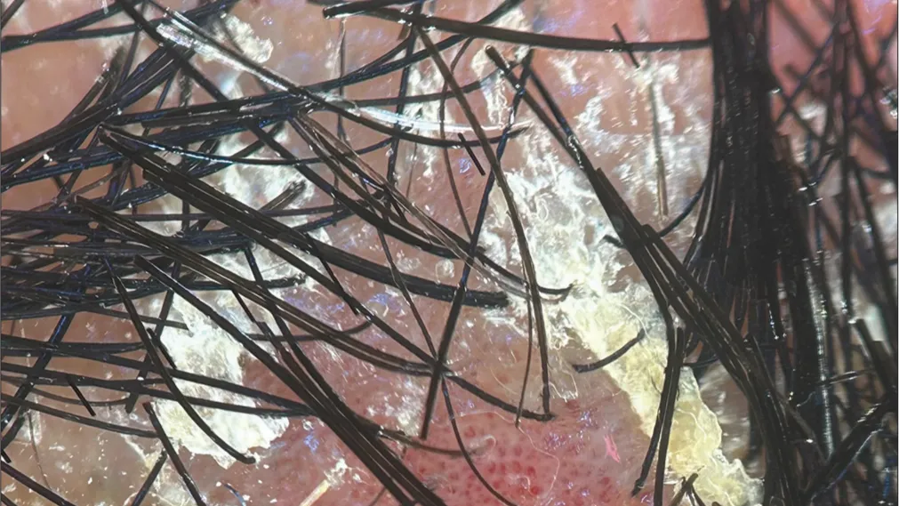
The most pathognomonic feature is the presence of regularly distributed red dots. These represent the tips of dilated and elongated capillaries within the dermal papillae. As the condition advances, these dots may evolve into twisted, coil-like vascular loops. The high degree of symmetry in their distribution is a primary differentiator from other inflammatory scalp conditions.
2. Scaling Patterns: Silvery-White and Amorphous
Unlike the greasy, yellowish scales of seborrheic dermatitis, the scales in scalp psoriasis appear dry, amorphous, and silvery-white. Under high magnification, these scales are typically diffuse and may occasionally reveal Munro micro-abscesses, which appear as tiny yellowish-white pustules trapped within the keratin layer.
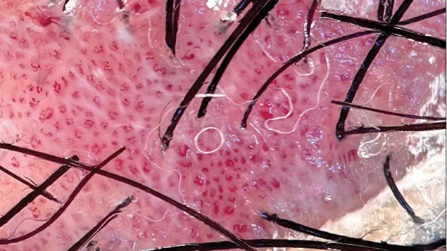
3. The Red Dot-In-Circle Sign
A specialized finding in early-stage scalp involvement is the red dot-in-circle sign. This consists of a central perifollicular capillary (the dot) surrounded by a concentric halo of white scaling. Recognizing this sign is essential for identifying psoriasis-related hair thinning before it becomes clinically apparent.
Differential Diagnosis: Psoriasis vs. Seborrheic Dermatitis
Distinguishing between these two conditions is the most frequent clinical requirement for dermoscopy. Use the following comparative framework for diagnostic calibration:
| Feature | Scalp Psoriasis | Seborrheic Dermatitis |
|---|---|---|
| Vascular Pattern | Uniform red dots and coil-like loops. | Atypical, thin, and branching (arborizing) vessels. |
| Scale Quality | Silvery-white, dry, and thick. | Yellowish, greasy, and thin. |
| Distribution | Regular and homogenous. | Patchy and irregular. |
| Auspitz Sign | Commonly observed after scale removal. | Absent. |
Advanced Visualization with IBOOLO Optical Systems
The precision of scalp psoriasis dermoscopy is significantly enhanced by the use of cross-polarized light. Devices like the IBOOLO DE-4100 Pro allow clinicians to toggle between modes to capture every detail:
- Polarized Mode: Best for observing the deeper vascular loops and blood vessels without the glare from the thick psoriatic scales.
- Non-Polarized Mode: Ideal for evaluating the surface texture and the exact silvery-white hue of the hyperkeratosis.
Furthermore, the ability to connect IBOOLO dermatoscopes to a smartphone enables teledermatology workflows, allowing for longitudinal monitoring of treatment efficacy through high-resolution image comparison.
Clinical Workflow for Scalp Examination
- Preparation: Ensure the scalp is free of heavy styling products. Gentle cleansing may be required to expose the underlying vascular patterns.
- Primary Scan: Use low magnification to assess the overall distribution of scaling and erythema.
- Detailed Analysis: Increase magnification to identify uniform red dots and determine the vascular morphology.
- Comparison: Contrast the findings with seborrheic dermatitis markers to rule out mimics.
Frequently Asked Questions
Can dermoscopy distinguish psoriasis from tinea capitis?
Yes. Tinea capitis (fungal infection) typically shows comma hairs, corkscrew hairs, or zigzag hairs, which are absent in scalp psoriasis. The vascular pattern of psoriasis is also much more organized.
Is immersion oil necessary for scalp examination?
While immersion oil can enhance transparency, modern polarized dermatoscopes like the IBOOLO series often provide sufficient clarity for vascular analysis without the mess of oil on the hair.
Recommended reading
How can I clean my dermoscopy after usage? – IBOOLO
Cleaning your dermoscopy after usage is important to prevent cross-contamination and infection. The cleaning method may vary depending on the type and model of your dermoscopy, so you should always follow the manufacturer’s instructions. However, some general steps are: • Turn off and disconnect your dermoscopy from any power source or device. • Wipe off...
Dermoscopy of Actinic Keratosis – IBOOLO
Actinic keratosis is a very common skin diseases. It is a major growing public health problem especially among older adults in white. A meta-analysis of observational studies reported by British Association of Dermatologists showed that the overall prevalence of actinic keratosis worldwide was 14%, with an estimated incidence of 1,928 cases per 100,000 peopl...
Affordable Polarized Light Dermoscopy Selection from Top China Products Supply - IBOOLO
As a leading affordable polarized light dermoscopy products supply in China, over 11+ years experience allows us to packed professional grade viewing into easy-to-carry mobile devices.


