


Article
Dermoscopy of Seborrheic Keratosis Dermatitis
Seborrheic keratosis dermatitis is a common skin hyperplasia. It is often mistaken for a disease such as skin cancer because of its appearance that looks like warts, precancerous skin growths, or skin cancer. Dermoscopy of seborrheic keratosis dermatitis is crucial to identify seborrhei keratosis from other types of skin diseases. What is Seborrheic Keratosis Dermatitis?Seborrheic…
Dermoscopy of Seborrheic Keratosis: An Expert Guide to Early and Irritated Lesions | IBOOLO
Confidently diagnose seborrheic keratosis. IBOOLO expert on seborrheic keratosis dermoscopy reveals key features for identifying and differentiating early and irritated lesions from malignant tumours.
Dermoscopy of Seborrheic Keratosis: An Expert Guide to Early and Irritated Lesions
Seborrheic keratosis (SK) is one of the most common benign skin tumours, often presenting as a harmless, waxy growth. However, its varied appearance can lead to a diagnostic dilemma, especially when it is in an early or irritated state, mimicking more serious conditions like basal cell carcinoma (BCC) or melanoma. Dermoscopy of seborrheic keratosis is a non-invasive diagnostic technique that has revolutionised its clinical assessment. By magnifying subtle microstructures, dermoscopy provides the visual clues necessary to distinguish SK from malignant lesions, ensuring accurate diagnosis and preventing unnecessary biopsies. This comprehensive guide will explore the classic dermoscopic features, delve into the complexities of irritated seborrheic keratosis dermoscopy and early seborrheic keratosis dermoscopy, and provide the critical knowledge needed to master this essential diagnostic skill.
The Classic Dermoscopic Features of Seborrheic Keratosis
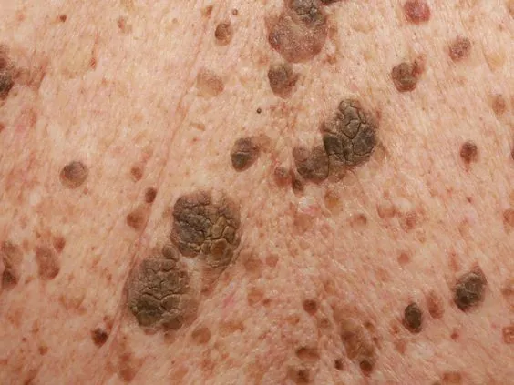
The diagnosis of a typical seborrheic keratosis lesion is often straightforward under dermoscopy. The classic signs are a result of the lesion's benign nature and are rarely seen in malignant tumors. Recognizing these features is the foundation of seborrheic keratosis dermoscopy.
1. Milia-like Cysts and Comedo-like Openings
These two features are the most reliable indicators of seborrheic keratosis. Milia-like cysts appear as small, round, white or yellowish structures embedded within the lesion. They are essentially tiny keratin cysts, reflecting the benign nature of the tumor. Comedo-like openings are dark, round, or oval-shaped structures that resemble the clogged pores or follicles of a comedone. These openings represent invaginations of the epidermis and are a hallmark of seborrheic keratosis. The presence of both features is a powerful confirmation of the diagnosis.
2. The "Pasted-On" Appearance and Fingerprint Structures
Under dermoscopy, the lesion often appears as though it has been "pasted" onto the skin, a distinct visual sign of its superficial, benign nature. This is a result of the sharp, well-demarcated border of the lesion. Within this border, some seborrheic keratoses have an intricate surface pattern that resembles fingerprints. This pattern is a series of parallel lines or a swirling, gyrate arrangement of ridges and grooves. These structures are a key sign in seborrheic keratosis dermoscopy and are a direct result of the lesion's acanthosis (thickening of the epidermis).
Navigating the Complexities: Irritated and Early Lesions
While a typical seborrheic keratosis is easy to identify, its presentation can be complicated by irritation or its early stage of development. This is where the true value of dermoscopy comes in, as it allows for the differentiation of these subtle variations from more serious skin cancers.
Irritated Seborrheic Keratosis Dermoscopy
When an SK becomes inflamed from friction or scratching, its dermoscopic features can change, leading to a diagnostic challenge. The inflammatory response can mask the classic signs and introduce new, potentially misleading features. However, by knowing what to look for, a clinician can confidently make a diagnosis.
- Erythema and Exudation: The most obvious sign of irritation is a background of diffuse erythema (redness). The lesion may also show signs of surface erosion or a central crust due to inflammation.
- Vascular Patterns: This is the most critical feature in irritated seborrheic keratosis dermoscopy. The inflammation can cause a proliferation of new blood vessels. These vessels typically appear as short, linear, or dotted structures, which are distinct from the chaotic, polymorphic vessels of melanoma or the arborizing vessels of BCC. The appearance of seborrheic keratosis dermoscopy vessels is a key diagnostic point—they are often uniform and regularly distributed, reflecting a benign process.
- Masked Features: The inflammation can obscure milia-like cysts or comedo-like openings, making the diagnosis more challenging. The clinician must look for any remaining classic signs on the periphery of the lesion.
Early Seborrheic Keratosis Dermoscopy
Diagnosing an early seborrheic keratosis is crucial for patient reassurance and management. These lesions are small and often lack the classic features, requiring a keen eye to spot the subtle, developing clues. Early seborrheic keratosis dermoscopy focuses on identifying these nascent characteristics.
- Subtle Pigmentation: Early lesions may show a slight, uniform, and light-brown pigmentation without the distinct pigment network seen in melanocytic nevi.
- Emerging Structures: The "pasted-on" appearance may be subtle. Look for a faint, emerging fingerprint pattern or early signs of a comedo-like opening, which may appear as a single, tiny black dot.
- Lack of Malignant Signs: The most important rule in diagnosing an early lesion is the absence of any signs of malignancy, such as irregular borders, chaotic pigmentation, or atypical vessels. The overall symmetry and organized pattern, however subtle, point towards a benign nature.
Differential Diagnosis: A Critical Comparison
The main reason for using dermoscopy for seborrheic keratosis is to differentiate it from malignant skin cancers. A side-by-side comparison of their dermoscopic features provides the clarity needed for a confident diagnosis.
Vs. Basal Cell Carcinoma (BCC)
BCC is the most common mimic of SK. Both can appear as raised, translucent, or pigmented nodules. The vascular patterns are the most reliable way to tell them apart. BCC is defined by arborizing vessels—large, branching blood vessels that look like tree limbs. These vessels are never seen in seborrheic keratosis. While SK can have some short, linear vessels (especially when irritated), they are not arborizing and are often accompanied by other classic SK features.
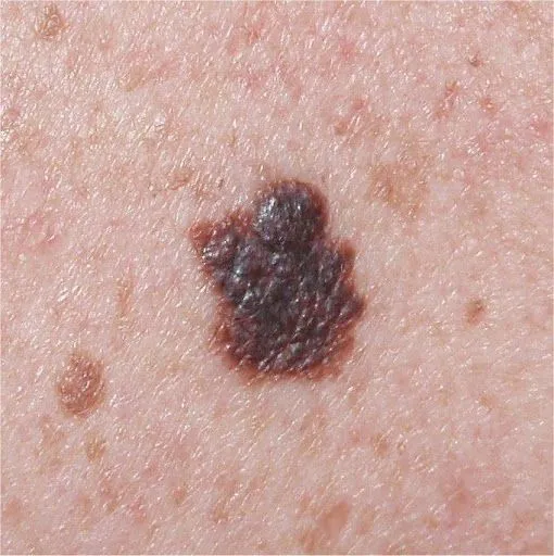
Vs. Melanoma
Pigmented seborrheic keratosis can be mistaken for melanoma. However, the dermoscopic features are fundamentally different. Melanoma is characterized by a lack of organization—asymmetry, irregular pigment networks, regression structures (scarring), and chaotic, polymorphic vessels. Seborrheic keratosis, by contrast, is a model of organization and benignity. It is defined by its symmetry, well-defined borders, and highly specific, organized structures like milia-like cysts and fingerprint patterns. The presence of these classic features effectively rules out melanoma.
The Clinical Workflow: Best Practices for Dermoscopy
Performing a dermoscopic examination of seborrheic keratosis requires a systematic approach to ensure all key features are evaluated.
- Patient History and Initial Assessment: Take a thorough history, focusing on the lesion's growth rate and any changes. Observe the lesion with the naked eye to note its color, elevation, and texture.
- Dermoscopic Examination: Use a dermoscope with both non-polarized and polarized light. Non-polarized light is best for observing the superficial features like comedo-like openings. Polarized light helps to visualize the deeper structures and vessels more clearly by eliminating surface glare, which is crucial for identifying seborrheic keratosis dermoscopy vessels in irritated lesions.
- Systematic Evaluation: Methodically scan the entire lesion. Look for a combination of classic features, paying close attention to the vessels. In irritated lesions, look for remnants of the classic features. In early lesions, look for the developing patterns.
- Documentation: Capture and save high-resolution images. This provides a clear record for future follow-up and for comparison with other lesions.
The Value of Dermoscopy
The ability to confidently diagnose seborrheic keratosis is a cornerstone of dermatological practice. Dermoscopy transforms a challenging visual assessment into a precise and reliable diagnostic process. By mastering the classic features, recognizing the subtleties of early seborrheic keratosis dermoscopy, and understanding the inflammatory changes in irritated seborrheic keratosis dermoscopy, clinicians can provide superior care. This non-invasive technique not only prevents unnecessary biopsies but also empowers both the clinician and the patient with a clear understanding of the lesion's benign nature. In a world where every suspicious mole is a concern, dermoscopy provides the clarity and confidence needed to make a life-saving distinction.
Recommended reading
Can dermoscopy with an electronic dermatoscope detect cancer?
Clinical studies validate that quality electronic dermatoscopes allow users to visually detect many early signs of skin cancer development with accuracy approaching in-person expert analysis. Features like asymmetry, border irregularity, evolving diameter, new colors, etc. can be recognized using an electronic dermatoscope. So combining vigilant self-checks with an electronic dermatoscope s photo documentation capabilities greatly aids early stage non-melanoma and melanoma detection.
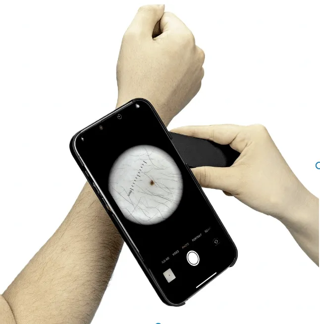
portable Dermoscopy power
Traditional dermatoscopes tend to be bulky desktop devices restricted to clinical settings. In contrast, today s handheld dermatoscopes enable portable dermoscopy anywhere. powered by batteries, these mobile phone dermatoscopes and compact handheld dermatoscopes free users from requiring an electrical outlet. Their lightweight, pocket-sized design allows easy whole-body examination in good lighting with a handheld dermatoscope.
Who Can perform Dermoscopy?
The powerful magnification and lighting capabilities dermatoscopes offer provide beneficial visual data for everyone to understand the current state of their skin and track changes over time. However, specialized medical training is typically required to analyze dermoscopy images and determine if biopsies or treatment are necessary. Dermatologists have this expertise.
Seborrheic keratosis dermatitis is a common skin hyperplasia. It is often mistaken for a disease such as skin cancer because of its appearance that looks like warts, precancerous skin growths, or skin cancer. Dermoscopy of seborrheic keratosis dermatitis is crucial to identify seborrhei keratosis from other types of skin diseases.
What is Seborrheic Keratosis Dermatitis?
Seborrheic keratosis dermatitis is a kind of benign epidermal hyperplasia caused by keratinocyte hyperplasia. Seborrheic keratosis dermatitis is a type of non-cancerous benign of skin disease. Seborrheic keratosis dermatitis is harmless.
Seborrheic keratosis dermatitis is known as senile warts, senile spots, also known as basal cell papilloma. Because it mainly occurs in adults over the age of 40, it often appears as people grow older.
What are the Clinical Feature of Seborrheic Keratosis Dermatitis?
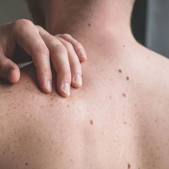
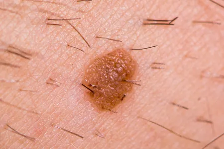
Seborrheic keratosis dermatitis is painless and it usually appears brown, black, or light tan. Its growth appears waxy or scaly and is slightly raised. They can gradually appear on various parts of the body, mostly on the face, neck, chest, or back.
Why is It Necessary to Use a Dermoscopy of Seborrheic Keratosis Dermatitis?
Dermatoscope is a non-invasive technique that allows dermatologists to closely examine seborrheic keratosis dermatitis more accurately and precisely. Especially dermoscopy of seborrheic keratosis dermatitis greatly enhance the vision of some locations that hard-to-reach by naked eyes, such as details in lesions. Dermatoscope magnifys and brightens shapes and structures of lesions. Dermoscopy of seborrheic keratosis dermatitis increases the confidence of doctors and patients about the skin disease and avoids unnecessary anxiety and treatment. So it is really necessary to use a dermoscopy of seborrheic keratosis.
Typical Features of Dermoscopy of Seborrheic Keratosis Dermatitis
dermoscopy plays a crucial role in identifying seborrheic keratosis dermatitis. There are some typical features of dermoscopy of seborrheic keratosis dermatitis as below:
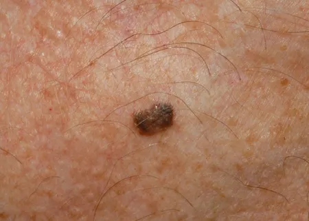
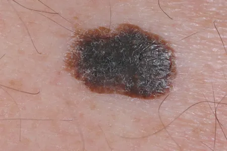
Special pattern: Typical “gyrigrain” or “fat-like” pattern.
Hair follicle openings: Visible hair follicle openings.
Structure: Edge ring structure, Light brown fingerprint-like parallel structures.
Prominent blood vessels: In some forms of seborrheic keratosis dermatitis, a halo of lobules surrounds tiny, hairpin-like capillaries.
Miliary cysts: These cysts may appear as small white stars or larger, yellowish turbidity.
Other features like: cracks/ridges, blue-gray balls, irregular crypts, weak or pseudo-network.
Dermoscopy of seborrheic keratosis dermatitis is very helpful and reliable for distinguish seborrheic keratosis dermatitis from other skin diseases.
How to differentiate between seborrheic keratosis dermatitis and melanoma?
Seborrheic kearatosis dermatitis will not transfer into melanoma. But both of seborrheic keratosis dermatitis and melanoma can be brown or black color, so the two can be easily be mistake from each other.
There are some important differences between seborrheic keratosis dermatitis and melanoma, from their numbers, appearances, locations causes, etc.
Comparison the apearances of seborrheic keratosis dermatitis and melanoma
Numbers: Seborrheic keratoses dermatitis: Seborrheic keratoses dermatitis often appear in numbers of two or more
Melanoma: Melanoma is usually appear in single.
Shapes: Seborrheic keratoses dermatitis: Seborrheic keratoses dermatitis usually shows round or oval shaped.
Melanoma: Irregular shape, asymmetry in shape is the typical features of melanoma.
Colors: Seborrheic keratoses dermatitis: Seborrheic keratoses dermatitis colors in light tan, brown, or black.
Melanoma: Melanoma is commonly display multiple colors like pink, red, white, blue or mixed color within the same one.
Size: Seborrheic keratosis dermatitis: Seborrheic keratosis dermatitis varies in size from very small to big, and its size will not changed as time goes on.
Melanoma: Melanoma is in bigger size than 1/4 inch, and its size will grow over time.
Surface: Seborrheic keratosis dermatitis: Seborrheic keratosis dermatitis has waxy or scaly surface, slightly elevated above the skin surface.
Melanoma: Melanoma tends to be smooth with blurred, ragged border.
Pain: Seborrheic keratosis dermatitis:Seborrheic keratosis dermatitis is painless
Melanoma: Some of melanoma feel hurt, while some of melanoma feel no any pain or discomfort.
Evolving: Seborrheic keratosis dermatitis:Seborrheic keratosis dermatitis maintains the same always.
Melanoma: Melanoma may looks different from its beginning, and it may change in its shape, size or color.
Comparison the locations of seborrheic keratosis dermatitis and melanoma
Seborrheic keratosis dermatitis: Seborrheic keratosis dermatitis mostly displays on the face, neck, chest, or back.
Melanoma: Melanoma Melanoma can appear in anywhere on the skin,mostly on chest, black, legs, arms, face, necks, and even eyes.
Comparison the causes of seborrheic keratosis dermatitis and melanoma
Causes: The primary risk factor for seborrheic keratoses is age. Other risk factors include: sunburn, skin irritation and friction, pregnancy, hormone therapy, some medications, genetic mutation, a family history of seborrheic keratosis
How is seborrheic keratosis dermatitis diagnosed?
To diagnose seborrheic keratosis dermatitis, skin doctors will get information from your family history of skin disease and take observation of it through a vision aiding tool called dermatoscope. Dermatoscope is a small handheld lighted medical microscope that allows a more precise and deeper view of skin diseases by high magnification and a powerful glare-free lighting system. If it is necessary, a biopsy will be needed for seborrheic keratosis.
People usually also take dermoscopy of seborrheic keratosis dermatitis for self-examination on skin. Any unusual findings or changes occur, have dermatologist checked for a further evaluation. Dermoscopy of seborrheic keratosis dermatitis plays a significant role in physical exam.
Application of dermoscopy of seborrheic keratosis dermatitis
A dermoscope is a main device used to examine skin diseases, like seborrheic keratosis dermatitis. In the diagnosis and treatment of seborrheic keratosis dermatitis, dermoscopy of seborrheic keratosis dermatitis is widely used in the following aspects:
Monitoring: For patients who have already been diagnosed with seborrheic keratosis dermatitis, dermoscopy can be used to monitor the whole process as time goes on. If any changes happen, a further step should be taken.
Feedback: Skin doctor can compare images from dermoscopy of seborrheic keratosis dermatitis over different times to assess the effectiveness of the treatment of seborrheic keratosis dermatitis and then decide if the treatment needs to be adjusted or not.
Treatment aid: When treating seborrheic keratosis dermatitis, images can be clearly and precisely showed by dermoscopy of seborrheic keratosis dermatitis. It greatly enhanced the patience of skin doctors and patients.
Seborrheic keratosis dermatitis is a harmless skin disease which will not cause skin cancer. But skin doctors should have it accurately diagnosed by dermoscope. Dermoscopy of seborrheic keratosis dermatitis is very important to differentiate seborrheic keratosis dermatitis from other skin diseases. Hence, skin doctors can remove seborrheic keratoss surely and safely for some aesthetic reasons.
It is vital to develop the habit of use of dermoscopy of seborrheic keratosis dermatitis. In addition, paying more attention to a regular skin examination is also necessary in our daily life. People should remain vigilant at all time to keep the health of the skin.


