IBOOLO Dermatoscope Light – IBOOLO

Shenzhen Iboolo Optics Co.Ltd was founded in 2012, whose major products are Macro lens, Woods Lamp, Dermatoscope and Microscope. Our business is currently merging R&D, production, and marketing after years of developments.

Article

IBOOLO Dermatoscope Light

Different IBOOLO dermatoscope models come with different lighting modes. The IBOOLO mini dermatoscopes DE-300 and DE-400 only have two lighting modes: white polarised and white non-polarised light. The handheld dermatoscopes DE-3100 and DE-4100, however, offer a more diverse range of lighting modes. In addition to the two aforementioned modes, they also feature white amber polarised…

Different IBOOLO dermatoscope models come with different lighting modes. The IBOOLO mini dermatoscopes DE-300 and DE-400 only have two lighting modes: white polarised and white non-polarised light. The handheld dermatoscopes DE-3100 and DE-4100, however, offer a more diverse range of lighting modes. In addition to the two aforementioned modes, they also feature white amber polarised light, amber light, and white amber non-polarised light, which can meet a variety of usage requirements.
What is Polarised Light and Non-Polarised Light in Dermatoscopy?
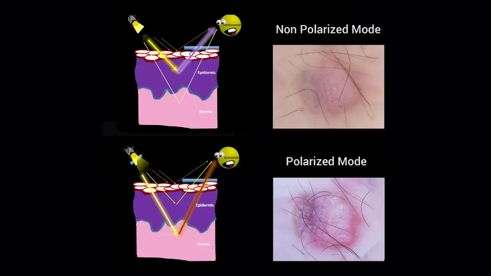
Polarised and Non-polarised light are the two basic modes of illumination used in dermoscopy. Non-Polarised contact dermoscopy requires the use of a contact solution to reduce reflections in order to examine superficial skin structures from the epidermis to the epidermal-dermal junction. Polarised dermoscopy, on the other hand, reduces reflections by blocking reflected light from the surface of the skin through cross-polarization techniques to better visualize deeper skin structures such as the epidermal-dermal junction and the superficial dermis. Polarised light is critical in dermoscopy because it enhances the visibility of certain skin features and is one of the most important modes of illumination.

Polarised VS. Non-polarised


Why is Polarised Light Important in Dermatoscopy?
All IBOOLO dermatoscope models except the first generation DE-200 are paired with polarised and non-polarised light. Polarised light offers several advantages in dermatoscopy. It doesn’t require direct skin contact or a contact liquid, making it suitable for examining infected or tender areas. The lack of pressure from non-contact polarised dermatoscopy preserves the skin’s geometry and circulation, aiding in the detection of small vascular structures. Polarised light is particularly effective in visualising deeper skin layers, such as the dermo-epidermal junction and superficial dermis. It can highlight structures like dermal vessels, pink/red colours, and white shiny structures, which are often associated with conditions like basal cell carcinoma. Additionally, polarised light can increase the sensitivity for detecting amelanotic melanoma or structure-poor melanoma, as it can better visualise blood vessels, vascular blush, and white shiny lines.
How Does IBOOLO’s Polarised Light Mode Work?
IBOOLO’s polarised light mode uses cross-polarisation filters. The source light is initially polarised with the first filter, and the reflected light from the skin is blocked by the second filter. This technique significantly reduces glare and allows for better visualisation of deeper skin structures. In this mode, the dermatoscope can be used in both contact and non-contact ways. When in contact with the skin, a contact liquid is not mandatory, which is convenient for examining various skin lesions. The polarised light mode is especially useful for viewing structures in the deeper layers of the skin and can help in the diagnosis of conditions like basal cell carcinoma, dermatofibroma, and some cases of melanoma.

IBOOLO Dermatoscope
IBOOLO Dermatoscope


Can IBOOLO’s Amber Polarised Light Enhance Pigment Contrast?
Yes, IBOOLO’s Amber polarised light can enhance the visualisation of pigmented structures within skin lesions. The Amber polarised light boosts the contrast of skin pigments, making it easier to observe features like the blue-white veil and Milia-like cysts. This lighting mode combines the benefits of polarised light in reducing glare and visualising deeper structures with the enhanced pigment contrast provided by the Amber wavelength. This combination is particularly useful for evaluating pigmented lesions and can improve the diagnostic accuracy for conditions like melanoma.
Is UV Light Useful in IBOOLO Dermatoscopy?
IBOOLO incorporates UV light (365nm) into its dermatoscope, offering Wood-mode capabilities. UV light in dermatoscopy can reveal fluorescing features that are not easily visible under visible light. It is especially useful for assessing the efficacy of acne treatment by visualising porphyrins produced by acne-causing bacteria. Furthermore, UV light can help discover exciting presentations of pigmented, vascular, or inflammatory lesions. It is also valuable in visualising bacterial and fungal infections on the skin, nails, or mucosa. With IBOOLO’s UV light mode, practitioners can gain additional diagnostic insights and evaluate a broader range of skin conditions. Currently only the DE-3100 PRO and DE-4100 PRO have built-in UV light illumination. However, in the upcoming DE-500 and DE-5100, the regular versions will also have UV light capability.

IBOOLO 365nm UV Light


How Can IBOOLO’s Various Lighting Modes Improve Diagnostic Accuracy?
IBOOLO’s advanced lighting modes—polarized (white/amber), non-polarized, and UV—offer a multi-layered approach to skin analysis. By understanding how each mode interacts with skin structures, clinicians can unlock deeper diagnostic insights, from early melanoma detection to infectious disease management. For example, the combination of polarised and non-polarised light can reveal both superficial and deeper structures, enhancing diagnostic confidence. The enhanced pigment contrast offered by amber polarised light aids in the early detection of pigmented lesions, while UV light provides additional insights into conditions like acne and infections. These varied lighting modes allow for a more thorough evaluation of skin lesions, enabling healthcare professionals to make more accurate diagnoses and develop more effective treatment plans.

Tags

Share this article

0

No products in the cart.

Have questions on gear or your order?

Our Gear Guides are here to help! Get personal advice from pro creatives

Name
Subject
Email address
How can we help?

Instant Answers

How can dermoscopy images be captured?

Dermoscopy images can be captured and stored in different ways, such as: • Using a smartphone or tablet with dermoscopic adapter, which consisted in the package.• Using a digital camera

Dermoscopy images can be captured and stored in different ways, such as:

• Using a smartphone or tablet with dermoscopic adapter, which consisted in the package.
• Using a digital camera with dermoscopic adapter, there’s 49mm screw size camera adapter available to order now.

Compatible phone/tablet models:
All iPhone models, 95% Android phones, 90% tablet. For phone/tablet size in 5.25-14mm

Compatible camera models:
All camera with built 49mm filter screw, such as Canon EOS 70D, 80D, 90D; Canon EOS R7, R10, R50, R100; Canon M100, M200, M50, Mark II; Canon G7X Mark III, Sony ZV-1

How can I connect my phone to my dermatoscope?

There’s universal phone adapter for all our dermoscopes. Please check the installation procedure bellow or watch operation guide. Smartphone Connector (1) Place phone adapter screw in the center of smartphone’s

There’s universal phone adapter for all our dermoscopes. Please check the installation procedure bellow or watch operation guide.

Smartphone Connector

(1) Place phone adapter screw in the center of smartphone’s main camera.
(2) Screw magnet attachment on phone adapter.
(3) Put dermoscope’s back ring and magnet attachment together

Take The Best Images

You need to adjust the focus ring after the dermoscpe connected on smartphone to get the best images.

How can I clean my dermoscopy after usage?

Cleaning your dermoscopy after usage is important to prevent cross-contamination and infection. The cleaning method may vary depending on the type and model of your dermoscopy, so you should always

Cleaning your dermoscopy after usage is important to prevent cross-contamination and infection. The cleaning method may vary depending on the type and model of your dermoscopy, so you should always follow the manufacturer’s instructions. However, some general steps are:

• Turn off and disconnect your dermoscopy from any power source or device.

• Wipe off any visible dirt or debris from the dermoscopy with a soft cloth or tissue.

• Disinfect the dermoscopy with an alcohol-based wipe or spray, or a disinfectant solution recommended by the manufacturer. Make sure to cover all surfaces, especially the lens and contact plate.

• Let the dermoscopy air dry completely before storing it in a clean and dry place.

• Do not use abrasive or corrosive cleaners, solvents, or detergents that may damage the dermoscopy.

• Do not immerse the dermoscopy in water or any liquid, unless it is waterproof and designed for immersion.

You should clean your dermoscopy after each use, or at least once a day if you use it frequently. You should also check your dermoscopy regularly for any signs of damage or malfunction, and contact the manufacturer or service provider if needed.

Polarized VS Non-polarized Dermoscopy

A dermoscopy is a device that allows the examination of skin lesions with magnificationand illumination. By revealing subsurface structures and patterns that are not visible tothe naked eye. It can

A dermoscopy is a device that allows the examination of skin lesions with magnificationand illumination. By revealing subsurface structures and patterns that are not visible tothe naked eye. It can improve the diagnose accuracy of skin lesions, such as melanoma,basal cell carcinoma, seborrheic keratosis, etc.

There are two main types of dermoscopy: Non polarized and polarized dermoscopy.We’ve fitted most of our dermoscopys with polarized and non-polarized light. They canbe used in multiple skin structures.

Non-polarized contact Mode

In non-polarized mode, the instrument can provide information about the superficialskin structures, such as milia-like cysts, comedo-like openings, and pigment in theepidemis.

The dermoscopy requires applying a liquid such as mineral oil or alcohol to the skin andplacing the lens in contact with the skin. This reduces surface reflection and enhancesthe view of subsurface structures.

Image with non-polarized light (DE-3100)

Polarized contact Mode

In polarized mode, the instrument allows for visualization for deeper skin structures,such as blood vessels, collagen, and pigment in the dermis.

The dermoscopy does not need to be in contact with the skin or use any liquid. Theirpolarized light can help to eliminate surface reflection and allow visualization ofvascular structures.

Image with polarized light (DE-3100)

Polarized non-contact Mode

The dermoscopy can also use polarized light to examine the skin without direct contact.

In polarized non-contact mode, the instrument allows for examination infected areasand lesions that are painful for the patient, or the difficult to contact pigmented lesions,such as nails and narrow areas.

The contact plate should be removed in this mode, and it does not require applying aliquid to the skin. As it doesn’t require pressure or fluid application on the skin, it canalso avoid cross-contamination and infection risk.

Image in polarized non-contact mode (DE-3100)

How effectiveness is dermoscopy

Compared with visual inspection, the dermoscopy can be used to capture and store skin lesion photos, which play an important role in early skin cancer examination. The dermoscopy allows the

Compared with visual inspection, the dermoscopy can be used to capture and store skin lesion photos, which play an important role in early skin cancer examination.

The dermoscopy allows the examination of skin lesions with magnification and illumination. This can be greatly avoiding the factors that cause interference to visual detection. Such as lighting, skin color, hair and cosmetics.

Several studies have demonstrated that dermoscopy is useful in the identification of melanoma, when used by a trained professional.

It may improve the accuracy of clinical diagnosis by up to 35%
It may reduce the number of harmless lesions that are removed
In primary care, it may increase the referral of more worrisome lesions and reduce the referral of more trivial ones

A 2018 Cochrane meta-analysis published the accuracy of dermoscopy in the detection.

Table 1. Accuracy of dermoscopy in the detection of melanoma in adults
Detection Method Sensitivity, % Specificity, % Positive Likelihood Ratio NegativeLikelihood Ratio
Visual inspection alone (in person) 76 75 3.04 0.32
Dermoscopy with visual inspection (in person) 92 95 18 0.08
Image-based visual inspection alone (not in person) 47 42 0.81 1.3
Dermoscopy with image-based visual inspection (not in person) 81 82 4.5 0.23
ROC—receiver operating characteristic. *Estimated sensitivity calculated on the summary ROC curve at a fixed specificity of 80%.

As we can see, the dermoscope can improve the accuracy of diagnosis of skin lesions, especially melanoma.

Table 1. Accuracy of dermoscopy in the detection of melanoma in adults
Detection Method Sensitivity, % Specificity, % Positive Likelihood Ratio NegativeLikelihood Ratio
Visual inspection alone (in person) 79 77 3.4 0.27
Dermoscopy with visual inspection (in person) 93 99 93 0.07
Image-based visual inspection alone (not in person) 85 87 6.5 0.17
Dermoscopy with image-based visual inspection (not in person) 93 96 23 0.07
ROC—receiver operating characteristic. *Estimated sensitivity calculated on the summary ROC curve at a fixed specificity of 80%.

Characteristics of the dermatoscopic structure of the skin lesions include:

• Symmetry or asymmetry
• Homogeny/uniformity (sameness) or heterogeny (structural differences across the lesion)
• Distribution of pigment: brown lines, dots, clods and structureless areas
• Skin surface keratin: small white cysts, crypts, fissures
• Vascular morphology and pattern: regular or irregular
• Border of the lesion: fading, sharply cut off or radial streaks
• Presence of ulceration

There are specific dermoscopic patterns that aid in the diagnosis of the following pigmented skin lesions:

• Melanoma
• Moles (benign melanocytic naevus)
• Freckles (lentigos)
• Atypical naevi
• Blue naevi
• Seborrhoeic keratosis
• Pigmented basal cell carcinoma
• Haemangioma

We use cookies on this website to provide a better user experience. By continuing to browse the website, you are giving your consent to receive cookies on this site. For more details please read our Privacy Policy.

Hot Search Terms