IBOOLO Wood’s Lamp – IBOOLO

Shenzhen Iboolo Optics Co.Ltd is located in Shenzhen, Guangdong. With 11+ years of experience in camera lens industry. Our main products include Macro lens, Woods Lamp, Dermatoscope and Microscope. Our R&D team is composed by experienced experts, specialists and engineers, who can keep pace with the developments of new technology and are able to provide creative products to meet different requests from our clients.

Article

IBOOLO Wood’s Lamp

A Wood’s lamp, also known as a ultraviolet light lamp or black light, is a diagnostic tool that emits long-wave ultraviolet radiation at a wavelength of 320–400 nanometers. Invented by physicist Robert Williams Wood in 1903, this device leverages the unique fluorescent properties of certain biological and chemical substances. When exposed to UVA light, these…

A Wood’s lamp, also known as a ultraviolet light lamp or black light, is a diagnostic tool that emits long-wave ultraviolet radiation at a wavelength of 320–400 nanometers. Invented by physicist Robert Williams Wood in 1903, this device leverages the unique fluorescent properties of certain biological and chemical substances. When exposed to UVA light, these substances absorb the radiation and re-emit it as visible light of varying colors, depending on their composition.
Modern Wood’s lamps, such as the IBOOLO Wood’s Lamp, use filtered UV light to enhance specificity in medical diagnostics. Unlike regular UV lights, they minimize visible light interference, allowing clinicians to observe subtle fluorescence patterns on the skin. This non-invasive tool is widely used in dermatology to detect infections, pigment disorders, and metabolic abnormalities.

How to Choose the Model of IBOOLO Wood’s Lamp?
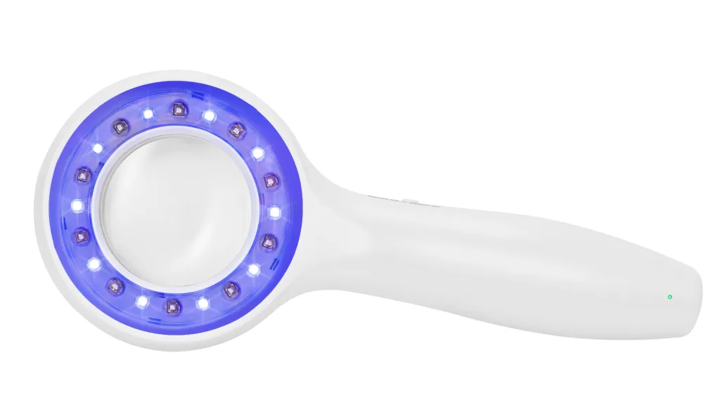
IBOOLO’s Wood Lamps are currently available in two models, the DE-215 and the DE-315, which differ in their focus and features. Equipped with a combination of 15 LED lights and 5 specialized 365nm UV beads, the IBOOLO DE-215 offers dual lighting modes tailored to meet your diverse inspection needs. The White Light Mode provides a crisp view of skin textures and surface details. The UV Mode, on the other hand, reveals hidden fungal and fluorescent reactions.
And IBOOLO DE-315 consists of 10 365nm and 10 405nm UV beads. So, the DE-315 is more focused on the UV mode than the DE-215. Users can select the appropriate UV wavelength according to their needs. 365nm is mainly used for highlighting skin pigmentation and superficial blood vessels, while 405nm is mainly used for ALA fluorescence imaging.

IBOOLO DE-315 Wood's Lamp
IBOOLO DE-315 Wood’s Lamp

Why is IBOOLO Wood’s Lamp Used?
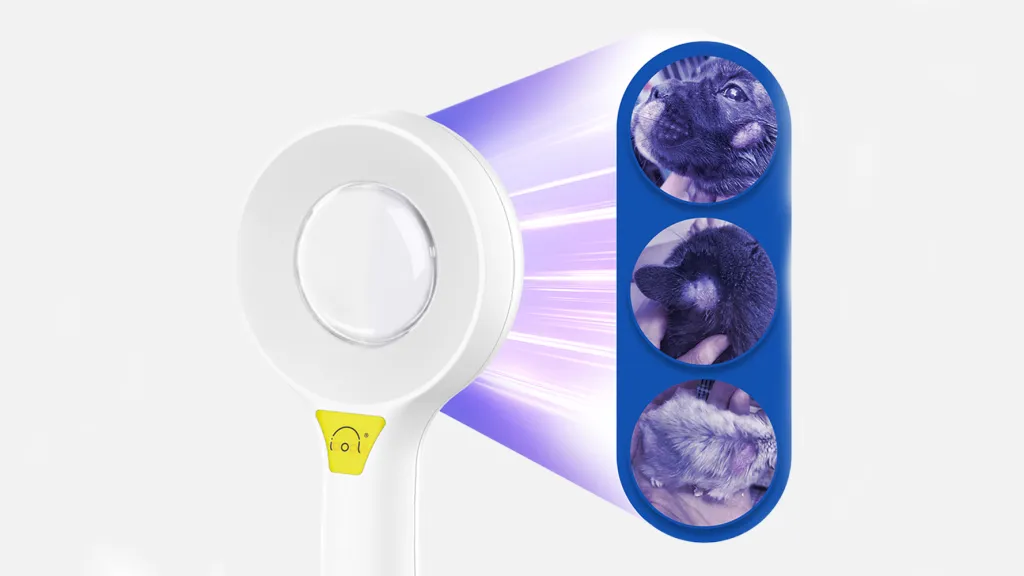
IBOOLO Wood’s Lamp is used for examining skin and hair under dark environment. It helps in identifying the extent of pigmented or depigmented patches and detecting fluorescence. This is possible because certain substances, such as collagen and porphyrins, absorb black light and emit it again at a longer wavelength in the visible spectrum. The lamp can highlight differences between hypo- or hyperpigmentations and reveal the accumulation of exogenous fluorophores, such as those produced by fungal and bacterial infections, or endogenous ones, as in the case of porphyrias.
How Does IBOOLO Wood’s Lamp Help in Observing Skin Diseases?
IBOOLO Wood’s Lamp can be used to observe various skin conditions. For pigmented disorders, it can determine whether pigmentation is epidermal or dermal. In cases of vitiligo, it can identify affected areas in light-skinned people. It is also useful in diagnosing conditions like erythrasma, tinea versicolor, Pityriasis versicolor, Malassezia folliculitis, Tinea capitis, head lice, scabies, acne, porphyria, and even the evenness of application of certain chemical peels. Each of these conditions presents specific fluorescence patterns under the lamp, aiding in accurate diagnosis.

IBOOLO Wood's Lamp
IBOOLO Wood’s Lamp


How is an IBOOLO Wood’s Lamp Skin Examination Performed?
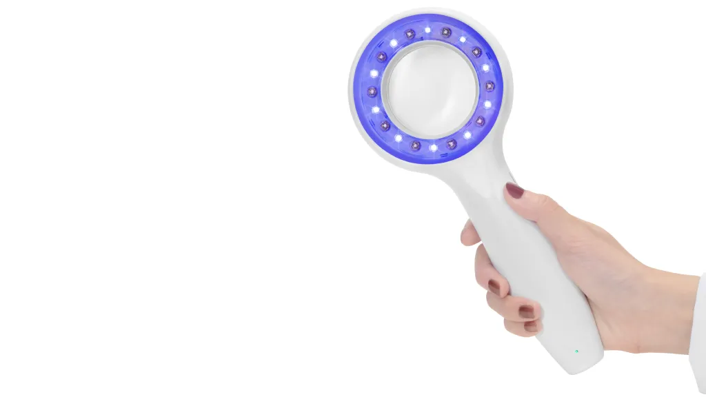
The Wood’s lamp examination process involves several steps. First, the skin to be examined should not have been recently washed or have had makeup, deodorant, or moisturizer applied, as these may error the results. However, it is important to emphasize that the skin should be clean before the examination. Secondly, too bright a light or environment can interfere with the fluorescent response of the Wood’s lamp. Therefore, please turn on the Wood’s lamp for observation in a dim environment or after using a light shield. The light should be about 10 – 30 centimeters away from the skin. The procedure is painless and safe.
Does IBOOLO Wood’s Lamp Have Any Harmful Effects?
The black light emitted by the IBOOLO Wood’s Lamp is generally harmless. It does not emit short – wavelength ultraviolet B radiation, so it does not cause sunburn or otherwise damage healthy skin. However, patients with extreme photosensitivity might develop a rash on skin exposed to black light. But Wood lamp examination is usually very brief and unlikely to cause problems even in very photosensitive patients. It is prudent to ask the patient to close their eyes when examining the face, especially for children, as their lenses lack the protective pigment found in adults, which absorbs UVA radiation, allowing it to reach the retina.

IBOOLO Woods Lamp Examination


Why Is Wood’s Lamp Better Than the Naked Eye and Other Devices?
When it comes to detecting skin conditions like vitiligo and fungal infections, Wood’s lamp offers distinct advantages over the naked eye and other diagnostic tools. The naked eye can miss subtle signs, especially in early stages or mild cases. Wood’s lamp, with its UV light, makes certain features glow, revealing what’s hidden to the naked eye. Compared to other devices, it’s non – invasive and doesn’t need skin samples. For vitiligo, it shows the sharp borders of pigment – free skin, helping determine the condition’s extent and depth. In fungal infections, it can highlight the presence of specific fungi that give off a characteristic glow, aiding quick and accurate identification. Its portability and ease of use also make it a practical choice for both quick checks and detailed exams in clinical settings.

Tags

Share this article

0

No products in the cart.

Have questions on gear or your order?

Our Gear Guides are here to help! Get personal advice from pro creatives

Name
Subject
Email address
How can we help?

Instant Answers

How can dermoscopy images be captured?

Dermoscopy images can be captured and stored in different ways, such as: • Using a smartphone or tablet with dermoscopic adapter, which consisted in the package.• Using a digital camera

Dermoscopy images can be captured and stored in different ways, such as:

• Using a smartphone or tablet with dermoscopic adapter, which consisted in the package.
• Using a digital camera with dermoscopic adapter, there’s 49mm screw size camera adapter available to order now.

Compatible phone/tablet models:
All iPhone models, 95% Android phones, 90% tablet. For phone/tablet size in 5.25-14mm

Compatible camera models:
All camera with built 49mm filter screw, such as Canon EOS 70D, 80D, 90D; Canon EOS R7, R10, R50, R100; Canon M100, M200, M50, Mark II; Canon G7X Mark III, Sony ZV-1

How can I connect my phone to my dermatoscope?

There’s universal phone adapter for all our dermoscopes. Please check the installation procedure bellow or watch operation guide. Smartphone Connector (1) Place phone adapter screw in the center of smartphone’s

There’s universal phone adapter for all our dermoscopes. Please check the installation procedure bellow or watch operation guide.

Smartphone Connector

(1) Place phone adapter screw in the center of smartphone’s main camera.
(2) Screw magnet attachment on phone adapter.
(3) Put dermoscope’s back ring and magnet attachment together

Take The Best Images

You need to adjust the focus ring after the dermoscpe connected on smartphone to get the best images.

How can I clean my dermoscopy after usage?

Cleaning your dermoscopy after usage is important to prevent cross-contamination and infection. The cleaning method may vary depending on the type and model of your dermoscopy, so you should always

Cleaning your dermoscopy after usage is important to prevent cross-contamination and infection. The cleaning method may vary depending on the type and model of your dermoscopy, so you should always follow the manufacturer’s instructions. However, some general steps are:

• Turn off and disconnect your dermoscopy from any power source or device.

• Wipe off any visible dirt or debris from the dermoscopy with a soft cloth or tissue.

• Disinfect the dermoscopy with an alcohol-based wipe or spray, or a disinfectant solution recommended by the manufacturer. Make sure to cover all surfaces, especially the lens and contact plate.

• Let the dermoscopy air dry completely before storing it in a clean and dry place.

• Do not use abrasive or corrosive cleaners, solvents, or detergents that may damage the dermoscopy.

• Do not immerse the dermoscopy in water or any liquid, unless it is waterproof and designed for immersion.

You should clean your dermoscopy after each use, or at least once a day if you use it frequently. You should also check your dermoscopy regularly for any signs of damage or malfunction, and contact the manufacturer or service provider if needed.

Polarized VS Non-polarized Dermoscopy

A dermoscopy is a device that allows the examination of skin lesions with magnificationand illumination. By revealing subsurface structures and patterns that are not visible tothe naked eye. It can

A dermoscopy is a device that allows the examination of skin lesions with magnificationand illumination. By revealing subsurface structures and patterns that are not visible tothe naked eye. It can improve the diagnose accuracy of skin lesions, such as melanoma,basal cell carcinoma, seborrheic keratosis, etc.

There are two main types of dermoscopy: Non polarized and polarized dermoscopy.We’ve fitted most of our dermoscopys with polarized and non-polarized light. They canbe used in multiple skin structures.

Non-polarized contact Mode

In non-polarized mode, the instrument can provide information about the superficialskin structures, such as milia-like cysts, comedo-like openings, and pigment in theepidemis.

The dermoscopy requires applying a liquid such as mineral oil or alcohol to the skin andplacing the lens in contact with the skin. This reduces surface reflection and enhancesthe view of subsurface structures.

Image with non-polarized light (DE-3100)

Polarized contact Mode

In polarized mode, the instrument allows for visualization for deeper skin structures,such as blood vessels, collagen, and pigment in the dermis.

The dermoscopy does not need to be in contact with the skin or use any liquid. Theirpolarized light can help to eliminate surface reflection and allow visualization ofvascular structures.

Image with polarized light (DE-3100)

Polarized non-contact Mode

The dermoscopy can also use polarized light to examine the skin without direct contact.

In polarized non-contact mode, the instrument allows for examination infected areasand lesions that are painful for the patient, or the difficult to contact pigmented lesions,such as nails and narrow areas.

The contact plate should be removed in this mode, and it does not require applying aliquid to the skin. As it doesn’t require pressure or fluid application on the skin, it canalso avoid cross-contamination and infection risk.

Image in polarized non-contact mode (DE-3100)

How effectiveness is dermoscopy

Compared with visual inspection, the dermoscopy can be used to capture and store skin lesion photos, which play an important role in early skin cancer examination. The dermoscopy allows the

Compared with visual inspection, the dermoscopy can be used to capture and store skin lesion photos, which play an important role in early skin cancer examination.

The dermoscopy allows the examination of skin lesions with magnification and illumination. This can be greatly avoiding the factors that cause interference to visual detection. Such as lighting, skin color, hair and cosmetics.

Several studies have demonstrated that dermoscopy is useful in the identification of melanoma, when used by a trained professional.

It may improve the accuracy of clinical diagnosis by up to 35%
It may reduce the number of harmless lesions that are removed
In primary care, it may increase the referral of more worrisome lesions and reduce the referral of more trivial ones

A 2018 Cochrane meta-analysis published the accuracy of dermoscopy in the detection.

Table 1. Accuracy of dermoscopy in the detection of melanoma in adults
Detection Method Sensitivity, % Specificity, % Positive Likelihood Ratio NegativeLikelihood Ratio
Visual inspection alone (in person) 76 75 3.04 0.32
Dermoscopy with visual inspection (in person) 92 95 18 0.08
Image-based visual inspection alone (not in person) 47 42 0.81 1.3
Dermoscopy with image-based visual inspection (not in person) 81 82 4.5 0.23
ROC—receiver operating characteristic. *Estimated sensitivity calculated on the summary ROC curve at a fixed specificity of 80%.

As we can see, the dermoscope can improve the accuracy of diagnosis of skin lesions, especially melanoma.

Table 1. Accuracy of dermoscopy in the detection of melanoma in adults
Detection Method Sensitivity, % Specificity, % Positive Likelihood Ratio NegativeLikelihood Ratio
Visual inspection alone (in person) 79 77 3.4 0.27
Dermoscopy with visual inspection (in person) 93 99 93 0.07
Image-based visual inspection alone (not in person) 85 87 6.5 0.17
Dermoscopy with image-based visual inspection (not in person) 93 96 23 0.07
ROC—receiver operating characteristic. *Estimated sensitivity calculated on the summary ROC curve at a fixed specificity of 80%.

Characteristics of the dermatoscopic structure of the skin lesions include:

• Symmetry or asymmetry
• Homogeny/uniformity (sameness) or heterogeny (structural differences across the lesion)
• Distribution of pigment: brown lines, dots, clods and structureless areas
• Skin surface keratin: small white cysts, crypts, fissures
• Vascular morphology and pattern: regular or irregular
• Border of the lesion: fading, sharply cut off or radial streaks
• Presence of ulceration

There are specific dermoscopic patterns that aid in the diagnosis of the following pigmented skin lesions:

• Melanoma
• Moles (benign melanocytic naevus)
• Freckles (lentigos)
• Atypical naevi
• Blue naevi
• Seborrhoeic keratosis
• Pigmented basal cell carcinoma
• Haemangioma

We use cookies on this website to provide a better user experience. By continuing to browse the website, you are giving your consent to receive cookies on this site. For more details please read our Privacy Policy.

Hot Search Terms