Skin Lesions Under IBOOLO DE-500 Dermoscope – IBOOLO

The manufacturer and supplier of camera lens, Shenzhen Iboolo Optics Co.Ltd has years of experience. In this industry, we have extensive R&D, manufacturing, and marketing experience.

Article

Skin Lesions Under IBOOLO DE-500 Dermoscope

The DE-500, launched in July 2025 as the latest model in the IBOOLO pocket dermatoscope series, represents a significant performance upgrade compared to previous devices in the same line. What’s been improved in the IBOOLO DE-500? Compared with the DE-400, the DE-500 offers three imaging modes—polarized, non-polarized, and UV—as well as three levels of brightness…

The DE-500, launched in July 2025 as the latest model in the IBOOLO pocket dermatoscope series, represents a significant performance upgrade compared to previous devices in the same line.

What’s been improved in the IBOOLO DE-500?

Compared with the DE-400, the DE-500 offers three imaging modes—polarized, non-polarized, and UV—as well as three levels of brightness adjustment. Its powerful lighting options make the DE-500 suitable for examining all types of skin lesions, and when combined with its advanced optical system, the device delivers excellent image quality.

In addition, the DE-500 uses a magnetic connection, which is more convenient than the threaded connection of the DE-400. The DE-500 also adopts an etched glass technique, whereas the DE-400 uses screen printing; etching provides clearer and more durable markings than screen printing.

What is the actual imaging performance of the IBOOLO DE-500?

Non-polarized light is mainly used to see the texture and details of the skin surface, focusing on the epidermal layer of the skin; polarized light can eliminate stray light, so that the user can clearly see the condition of the dermis layer of the skin; 365nm UV light can be used to detect fungal lesions as well as pigmentation deficiency diseases, such as vitiligo, psoriasis and so on.

The following skin lesions were all photographed with the DE-500 connected to a cell phone. According to the characteristics of different skin lesions, the user chose different lighting shots for observation. 

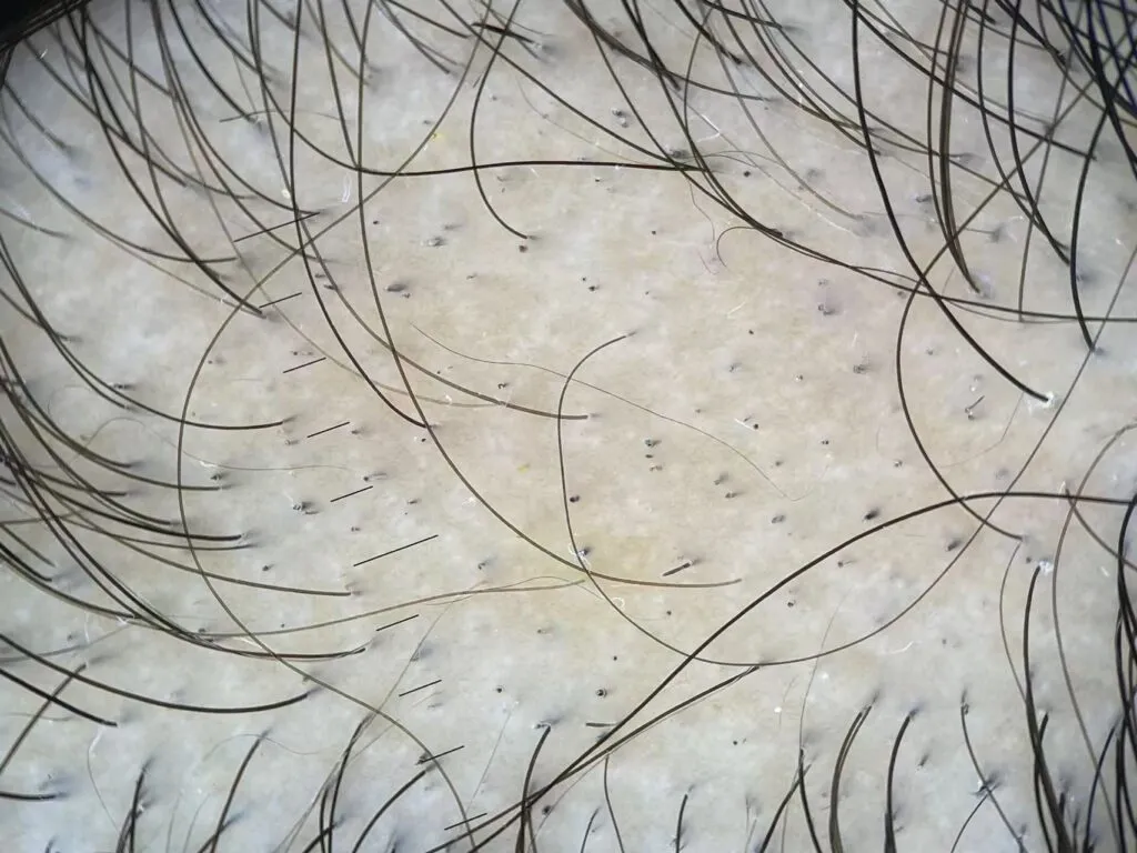
Alopecia Areata under IBOOLO DE-500 

Alopecia areata under dermoscopy, also known as trichoscopy, presents several characteristic findings that are useful in clinical practice. The most consistent features include yellow dots that represent keratin and sebum in dilated follicular openings, black dots corresponding to broken hairs at the scalp surface, and exclamation-mark hairs that are tapered proximally and broader distally, often located at the border of active patches. Short vellus hairs and cadaverized hair shafts are also frequently observed, and these signs together help differentiate alopecia areata from other types of nonscarring alopecia.

Alopecia areata IBOOLO DE-500 Dermatoscope
Alopecia Areata

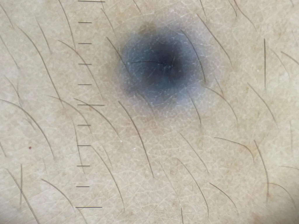
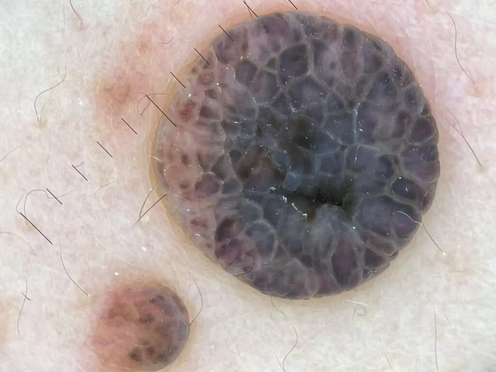
Blue Nevus under IBOOLO DE-500 

Blue nevus shows on dermoscopy a homogeneous, structureless, steel-blue to blue-gray pigmentation that corresponds to melanin located in the dermis. It usually lacks a pigment network, and the blue coloration is uniform across the lesion. In some cases, blue globules or dots may be visible, and the borders tend to be well defined. These features are important for distinguishing blue nevus from malignant melanoma or other pigmented lesions.

Blue Nevus IBOOLO DE-500  Dermatoscope
Blue Nevus

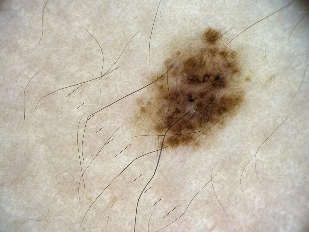
Congenital Melanocytic Nevus under IBOOLO DE-500 

Congenital melanocytic nevus demonstrates variable dermoscopic patterns depending on the size and anatomic site of the lesion. Small to medium congenital nevi often show a globular or reticular-globular pattern, while larger lesions may reveal a multicomponent pattern with heterogeneous pigmentation. On the face or scalp, perifollicular pigmentation and increased follicular openings can be noted. Documenting these dermoscopic features is important to establish a baseline for long-term monitoring.

Congenital Melanocytic Nevus IBOOLO DE-500 Dermatoscope
Congenital Melanocytic Nevus

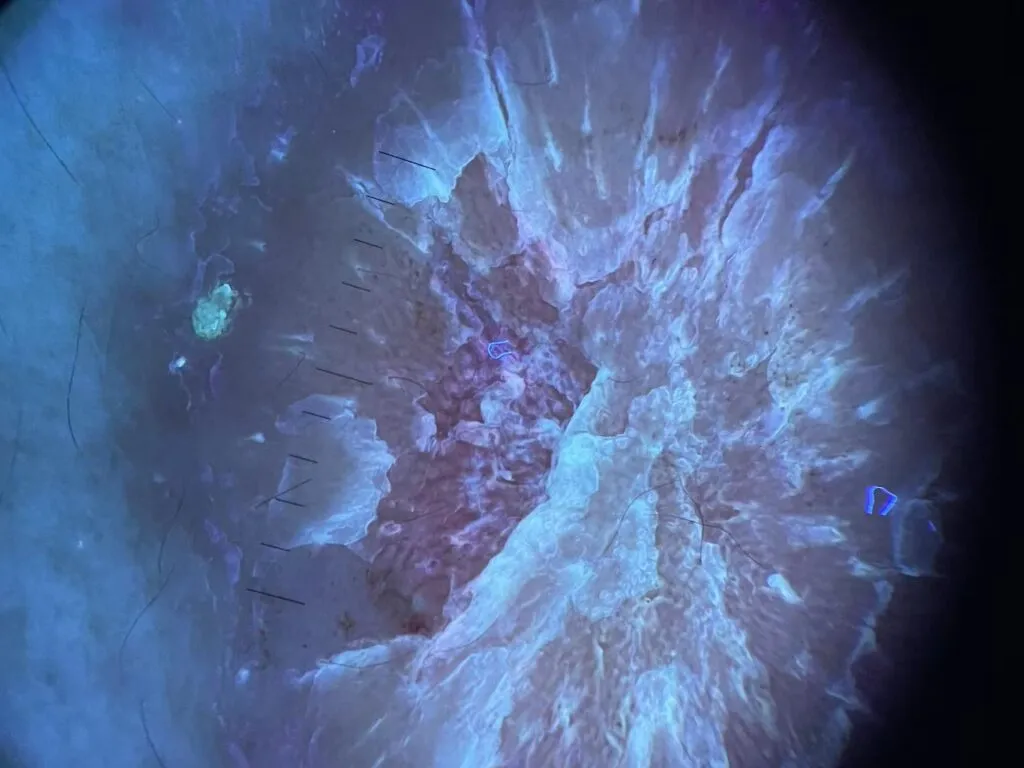
Epidermoid Cysts under IBOOLO DE-500 

Epidermoid cysts under dermoscopy are characterized by the presence of a central punctum or pore, often referred to as the “pore sign.” The cyst may also show homogeneous white-yellow or yellow-brown areas reflecting keratin content. When inflamed or ruptured, vascular structures such as arborizing or branching vessels and bluish or reddish discoloration may be seen. Identifying the punctum is particularly helpful for noninvasive diagnosis.

Epidermoid Cyst IBOOLO DE-500 Dermatoscope
Epidermoid Cyst

Intradermal Nevus under IBOOLO DE-500 

Intradermal nevus appears on dermoscopy as a lesion dominated by vascular rather than pigmented structures. Common findings include comma-shaped vessels and polymorphous vascular patterns on a skin-colored or slightly pigmented background. The lesion often lacks a pigment network or shows only faint pigmentation, which helps differentiate it from junctional or compound nevi and from early melanoma.

Intradermal Nevus
Intradermal Nevus

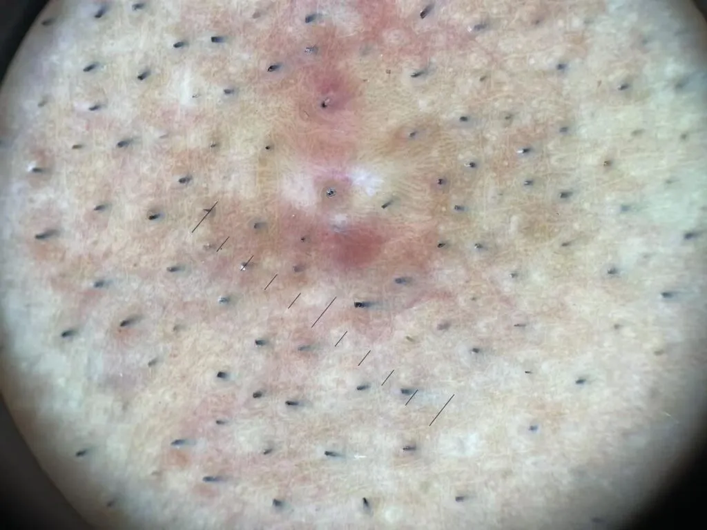
Psoriasis under IBOOLO DE-500 

Psoriasis exhibits on dermoscopy a distinctive vascular and scaling pattern. The most typical features are regularly distributed red dots that correspond to dilated capillaries in dermal papillae, set against a light-red background, with overlying white scales. In plaque psoriasis, red globules and twisted red loops may also appear, arranged in a symmetrical and uniform distribution. These features aid in distinguishing psoriasis from eczema or lichen planus.

Psoriasis DE-500 Dermatoscope
Psoriasis

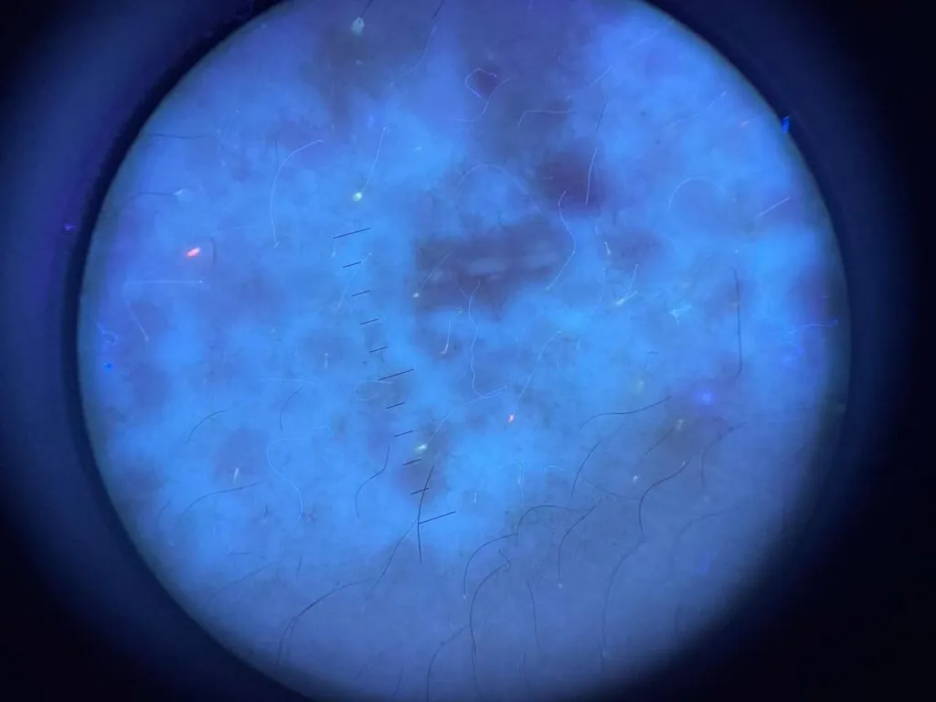
Vitiligo under IBOOLO DE-500 

Vitiligo under dermoscopy is characterized by structureless white areas that lack the normal pigment network. The borders of these depigmented patches are often sharply demarcated, and perifollicular pigmentation or a reversed pigment network may be observed at the margins. Other features include leukotrichia and starburst or micro-Koebner patterns, which may indicate active disease or ongoing repigmentation. These findings support clinical diagnosis and monitoring of disease activity.

Vitiligo
Vitiligo

Share this article

0

No products in the cart.

Ask

Have questions on gear or your order?

Our Gear Guides are here to help! Get personal advice from pro creatives

Name
Subject
Email address
How can we help?
Answers

Instant Answers

How can dermoscopy images be captured?

Dermoscopy images can be captured and stored in different ways, such as: • Using a smartphone or tablet with dermoscopic adapter, which consisted in the package.• Using a digital camera

Dermoscopy images can be captured and stored in different ways, such as:

• Using a smartphone or tablet with dermoscopic adapter, which consisted in the package.
• Using a digital camera with dermoscopic adapter, there’s 49mm screw size camera adapter available to order now.

Compatible phone/tablet models:
All iPhone models, 95% Android phones, 90% tablet. For phone/tablet size in 5.25-14mm

Compatible camera models:
All camera with built 49mm filter screw, such as Canon EOS 70D, 80D, 90D; Canon EOS R7, R10, R50, R100; Canon M100, M200, M50, Mark II; Canon G7X Mark III, Sony ZV-1

How can I connect my phone to my dermatoscope?

There’s universal phone adapter for all our dermoscopes. Please check the installation procedure bellow or watch operation guide. Smartphone Connector (1) Place phone adapter screw in the center of smartphone’s

There’s universal phone adapter for all our dermoscopes. Please check the installation procedure bellow or watch operation guide.

Smartphone Connector

(1) Place phone adapter screw in the center of smartphone’s main camera.
(2) Screw magnet attachment on phone adapter.
(3) Put dermoscope’s back ring and magnet attachment together

Take The Best Images

You need to adjust the focus ring after the dermoscpe connected on smartphone to get the best images.

How can I clean my dermoscopy after usage?

Cleaning your dermoscopy after usage is important to prevent cross-contamination and infection. The cleaning method may vary depending on the type and model of your dermoscopy, so you should always

Cleaning your dermoscopy after usage is important to prevent cross-contamination and infection. The cleaning method may vary depending on the type and model of your dermoscopy, so you should always follow the manufacturer’s instructions. However, some general steps are:

• Turn off and disconnect your dermoscopy from any power source or device.

• Wipe off any visible dirt or debris from the dermoscopy with a soft cloth or tissue.

• Disinfect the dermoscopy with an alcohol-based wipe or spray, or a disinfectant solution recommended by the manufacturer. Make sure to cover all surfaces, especially the lens and contact plate.

• Let the dermoscopy air dry completely before storing it in a clean and dry place.

• Do not use abrasive or corrosive cleaners, solvents, or detergents that may damage the dermoscopy.

• Do not immerse the dermoscopy in water or any liquid, unless it is waterproof and designed for immersion.

You should clean your dermoscopy after each use, or at least once a day if you use it frequently. You should also check your dermoscopy regularly for any signs of damage or malfunction, and contact the manufacturer or service provider if needed.

Polarized VS Non-polarized Dermoscopy

A dermoscopy is a device that allows the examination of skin lesions with magnificationand illumination. By revealing subsurface structures and patterns that are not visible tothe naked eye. It can

A dermoscopy is a device that allows the examination of skin lesions with magnificationand illumination. By revealing subsurface structures and patterns that are not visible tothe naked eye. It can improve the diagnose accuracy of skin lesions, such as melanoma,basal cell carcinoma, seborrheic keratosis, etc.

There are two main types of dermoscopy: Non polarized and polarized dermoscopy.We’ve fitted most of our dermoscopys with polarized and non-polarized light. They canbe used in multiple skin structures.

Non-polarized contact Mode

In non-polarized mode, the instrument can provide information about the superficialskin structures, such as milia-like cysts, comedo-like openings, and pigment in theepidemis.

The dermoscopy requires applying a liquid such as mineral oil or alcohol to the skin andplacing the lens in contact with the skin. This reduces surface reflection and enhancesthe view of subsurface structures.

Image with non-polarized light (DE-3100)

Polarized contact Mode

In polarized mode, the instrument allows for visualization for deeper skin structures,such as blood vessels, collagen, and pigment in the dermis.

The dermoscopy does not need to be in contact with the skin or use any liquid. Theirpolarized light can help to eliminate surface reflection and allow visualization ofvascular structures.

Image with polarized light (DE-3100)

Polarized non-contact Mode

The dermoscopy can also use polarized light to examine the skin without direct contact.

In polarized non-contact mode, the instrument allows for examination infected areasand lesions that are painful for the patient, or the difficult to contact pigmented lesions,such as nails and narrow areas.

The contact plate should be removed in this mode, and it does not require applying aliquid to the skin. As it doesn’t require pressure or fluid application on the skin, it canalso avoid cross-contamination and infection risk.

Image in polarized non-contact mode (DE-3100)

How effectiveness is dermoscopy

Compared with visual inspection, the dermoscopy can be used to capture and store skin lesion photos, which play an important role in early skin cancer examination. The dermoscopy allows the

Compared with visual inspection, the dermoscopy can be used to capture and store skin lesion photos, which play an important role in early skin cancer examination.

The dermoscopy allows the examination of skin lesions with magnification and illumination. This can be greatly avoiding the factors that cause interference to visual detection. Such as lighting, skin color, hair and cosmetics.

Several studies have demonstrated that dermoscopy is useful in the identification of melanoma, when used by a trained professional.

It may improve the accuracy of clinical diagnosis by up to 35%
It may reduce the number of harmless lesions that are removed
In primary care, it may increase the referral of more worrisome lesions and reduce the referral of more trivial ones

A 2018 Cochrane meta-analysis published the accuracy of dermoscopy in the detection.

Table 1. Accuracy of dermoscopy in the detection of melanoma in adults
Detection Method Sensitivity, % Specificity, % Positive Likelihood Ratio NegativeLikelihood Ratio
Visual inspection alone (in person) 76 75 3.04 0.32
Dermoscopy with visual inspection (in person) 92 95 18 0.08
Image-based visual inspection alone (not in person) 47 42 0.81 1.3
Dermoscopy with image-based visual inspection (not in person) 81 82 4.5 0.23
ROC—receiver operating characteristic. *Estimated sensitivity calculated on the summary ROC curve at a fixed specificity of 80%.

As we can see, the dermoscope can improve the accuracy of diagnosis of skin lesions, especially melanoma.

Table 1. Accuracy of dermoscopy in the detection of melanoma in adults
Detection Method Sensitivity, % Specificity, % Positive Likelihood Ratio NegativeLikelihood Ratio
Visual inspection alone (in person) 79 77 3.4 0.27
Dermoscopy with visual inspection (in person) 93 99 93 0.07
Image-based visual inspection alone (not in person) 85 87 6.5 0.17
Dermoscopy with image-based visual inspection (not in person) 93 96 23 0.07
ROC—receiver operating characteristic. *Estimated sensitivity calculated on the summary ROC curve at a fixed specificity of 80%.

Characteristics of the dermatoscopic structure of the skin lesions include:

• Symmetry or asymmetry
• Homogeny/uniformity (sameness) or heterogeny (structural differences across the lesion)
• Distribution of pigment: brown lines, dots, clods and structureless areas
• Skin surface keratin: small white cysts, crypts, fissures
• Vascular morphology and pattern: regular or irregular
• Border of the lesion: fading, sharply cut off or radial streaks
• Presence of ulceration

There are specific dermoscopic patterns that aid in the diagnosis of the following pigmented skin lesions:

• Melanoma
• Moles (benign melanocytic naevus)
• Freckles (lentigos)
• Atypical naevi
• Blue naevi
• Seborrhoeic keratosis
• Pigmented basal cell carcinoma
• Haemangioma

We use cookies on this website to provide a better user experience. By continuing to browse the website, you are giving your consent to receive cookies on this site. For more details please read our Privacy Policy.

Hot Search Terms