What are the Common Skin Cancers? – IBOOLO

Shenzhen Iboolo Optics Co.Ltd established in 2012, which is consist of experienced professional and executive team among our core technicians average camera lens trade research and production experience 11+ years. We are one of the top professional manufacturers and suppliers of Macro lens, Woods Lamp, Dermatoscope and Microscope.

Article

What are the Common Skin Cancers?

Skin cancer stands out as the most prevalent form of cancer globally, with millions of new diagnoses annually. Unlike many other cancers, skin cancer often presents visible signs, making early detection feasible. The American Cancer Society reports a steady rise in skin cancer incidence over recent decades. Among these, basal cell carcinoma (BCC), squamous cell…

Skin cancer stands out as the most prevalent form of cancer globally, with millions of new diagnoses annually. Unlike many other cancers, skin cancer often presents visible signs, making early detection feasible. The American Cancer Society reports a steady rise in skin cancer incidence over recent decades. Among these, basal cell carcinoma (BCC), squamous cell carcinoma (SCC), and melanoma are the most frequently encountered types. BCC is the most common, accounting for approximately 80% of all skin cancers. SCC follows, making up around 20%, while melanoma, though less common, is the most aggressive and deadly. Early detection and treatment are vital for favorable outcomes and reduced complications.
What is Basal Cell Carcinoma?
Basal cell carcinoma (BCC) originates from the basal cells of the interfollicular epidermis and/or hair follicle. It is the most common type of skin cancer, accounting for about 80% of all skin malignancies. BCC can arise anywhere on the body but is most commonly seen on sun-exposed areas such as the face, neck, and arms. It typically presents as a slow-growing, pearly or flesh-colored nodule or plaque, which may ulcerate and bleed. BCC rarely metastasizes but can cause significant local tissue destruction and disfigurement if left untreated.
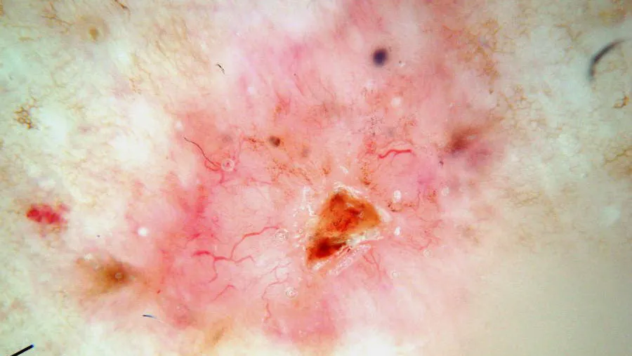
What are the Dermatoscopic Features of Basal Cell Carcinoma?
The dermoscopic features of BCC vary according to subtype. Pigmented BCC may exhibit features such as absence of pigment network, linear and arborizing telangiectasia, structureless or leaf-like areas on the periphery of the lesion, large blue-grey ovoid nests or blotches, multiple blue-grey globules, specks of brown and grey pigment, spoke wheel areas, and focal ulceration. Nonpigmented BCC typically shows bluish or whitish-pink stroma, asymmetrical branching serpentine vessels, focal ulceration, slight scaling, and white clues, particularly perpendicular white lines under polarized light and structureless roundish white or yellowish areas.

Basal cell carcinoma dermatoscope


What is Squamous Cell Carcinoma?
Squamous cell carcinoma (SCC) is another common type of skin cancer that arises from the keratinocytes of the epidermis. It is the second most common skin cancer, accounting for approximately 20% of all skin malignancies. SCC most often occurs on sun-exposed areas such as the face, ears, neck, hands, and forearms. It typically presents as a red, scaly plaque or nodule, which may ulcerate and have a crusted surface. SCC can metastasize, especially in high-risk populations such as immunocompromised individuals.
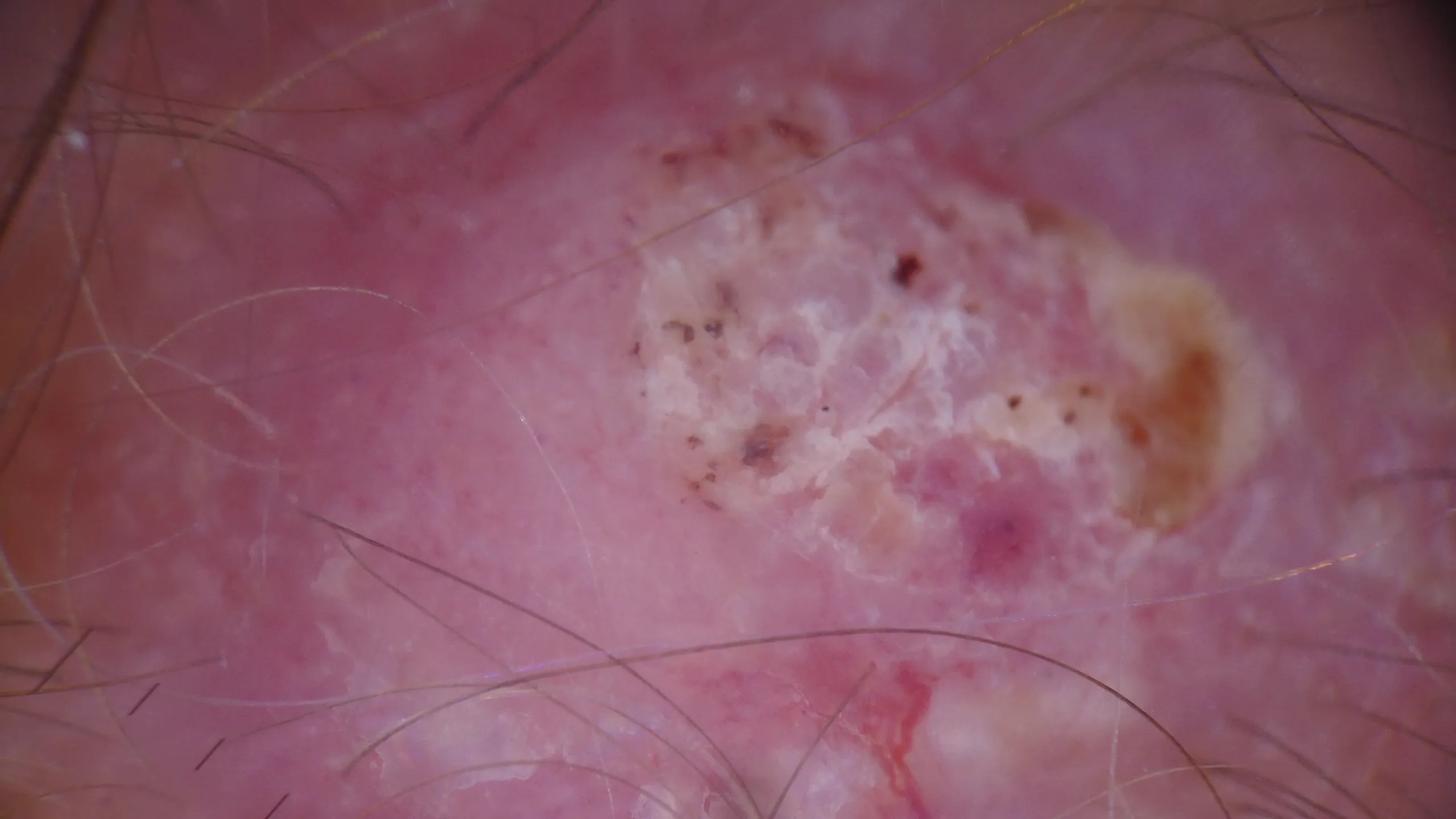
What are the Dermatoscopic Features of Squamous Cell Carcinoma?
The dermoscopic features of SCC may include well-defined borders, hyperkeratosis, scaling, erythema, and the presence of ulceration. Vascular patterns such as glomerular vessels or irregular dotted vessels may also be observed. SCC often shows asymmetry and pleomorphic vessels, which are important clues for diagnosis. Under polarized light dermoscopy, SCC may reveal additional features such as structureless areas, white patches, and keratin globules.

SCC dermatoscope

What is Melanoma?
Melanoma is a malignant tumor that arises from melanocytes, the cells responsible for producing melanin. It is less common than BCC and SCC but is the most aggressive and deadly form of skin cancer. Melanoma can occur anywhere on the body, including non-sun-exposed areas. It often presents as an asymmetric, irregularly bordered, multicolored mole or lesion. Early detection and treatment are critical for improving survival rates, as melanoma can metastasize rapidly to other parts of the body.
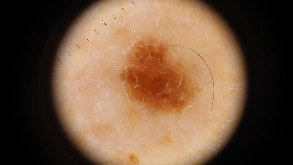
What are the Dermatoscopic Features of Melanoma?
The dermoscopic features of melanoma are diverse and can vary depending on the subtype and stage of the lesion. Common features include asymmetry, irregular borders, multiple colors (brown, black, blue, red, white), and specific structures such as atypical pigment networks, blue-black areas, and white or pink structureless areas. The presence of regression structures (e.g., blue-gray globules, black dots, and patches) and asymmetric follicular openings are also important indicators. In nodular melanoma, features such as a homogeneous blue-black or black color, irregular blotches, and multiple blue-black globules are often observed. For lentigo maligna melanoma, the presence of a broad and irregular pigment network, gray lines, pseudopodia, and multiple blue-gray globules at the periphery is characteristic. Acral lentiginous melanoma typically shows parallel ridge dermoscopic patterns, intersecting white lines, and multiple blue-black dots or globules.

melanoma dermatoscope


Is Dermatoscopy Effective for Skin Cancer?
Dermatoscopy is a non-invasive diagnostic technique that has revolutionized the detection and management of skin cancer. IBOOLO handheld dermatoscopes are capable of 10X magnification. They can reveal subtle details that are invisible to the naked eye. The IBOOLO DE-4100 has a variety of light modes, polarized light, unpolarized light, amber light, which can basically satisfy the observation of all skin lesions.
The ability to observe specific features like asymmetric borders, irregular pigmentation, and unusual vascular patterns helps in distinguishing benign lesions from malignant ones. Dermatoscopy not only improves diagnostic accuracy but also reduces the need for unnecessary biopsies by providing clearer information about the nature of a skin lesion.

Share this article

0

No products in the cart.

Have questions on gear or your order?

Our Gear Guides are here to help! Get personal advice from pro creatives

Name
Subject
Email address
How can we help?

Instant Answers

How can dermoscopy images be captured?

Dermoscopy images can be captured and stored in different ways, such as: • Using a smartphone or tablet with dermoscopic adapter, which consisted in the package.• Using a digital camera

Dermoscopy images can be captured and stored in different ways, such as:

• Using a smartphone or tablet with dermoscopic adapter, which consisted in the package.
• Using a digital camera with dermoscopic adapter, there’s 49mm screw size camera adapter available to order now.

Compatible phone/tablet models:
All iPhone models, 95% Android phones, 90% tablet. For phone/tablet size in 5.25-14mm

Compatible camera models:
All camera with built 49mm filter screw, such as Canon EOS 70D, 80D, 90D; Canon EOS R7, R10, R50, R100; Canon M100, M200, M50, Mark II; Canon G7X Mark III, Sony ZV-1

How can I connect my phone to my dermatoscope?

There’s universal phone adapter for all our dermoscopes. Please check the installation procedure bellow or watch operation guide. Smartphone Connector (1) Place phone adapter screw in the center of smartphone’s

There’s universal phone adapter for all our dermoscopes. Please check the installation procedure bellow or watch operation guide.

Smartphone Connector

(1) Place phone adapter screw in the center of smartphone’s main camera.
(2) Screw magnet attachment on phone adapter.
(3) Put dermoscope’s back ring and magnet attachment together

Take The Best Images

You need to adjust the focus ring after the dermoscpe connected on smartphone to get the best images.

How can I clean my dermoscopy after usage?

Cleaning your dermoscopy after usage is important to prevent cross-contamination and infection. The cleaning method may vary depending on the type and model of your dermoscopy, so you should always

Cleaning your dermoscopy after usage is important to prevent cross-contamination and infection. The cleaning method may vary depending on the type and model of your dermoscopy, so you should always follow the manufacturer’s instructions. However, some general steps are:

• Turn off and disconnect your dermoscopy from any power source or device.

• Wipe off any visible dirt or debris from the dermoscopy with a soft cloth or tissue.

• Disinfect the dermoscopy with an alcohol-based wipe or spray, or a disinfectant solution recommended by the manufacturer. Make sure to cover all surfaces, especially the lens and contact plate.

• Let the dermoscopy air dry completely before storing it in a clean and dry place.

• Do not use abrasive or corrosive cleaners, solvents, or detergents that may damage the dermoscopy.

• Do not immerse the dermoscopy in water or any liquid, unless it is waterproof and designed for immersion.

You should clean your dermoscopy after each use, or at least once a day if you use it frequently. You should also check your dermoscopy regularly for any signs of damage or malfunction, and contact the manufacturer or service provider if needed.

Polarized VS Non-polarized Dermoscopy

A dermoscopy is a device that allows the examination of skin lesions with magnificationand illumination. By revealing subsurface structures and patterns that are not visible tothe naked eye. It can

A dermoscopy is a device that allows the examination of skin lesions with magnificationand illumination. By revealing subsurface structures and patterns that are not visible tothe naked eye. It can improve the diagnose accuracy of skin lesions, such as melanoma,basal cell carcinoma, seborrheic keratosis, etc.

There are two main types of dermoscopy: Non polarized and polarized dermoscopy.We’ve fitted most of our dermoscopys with polarized and non-polarized light. They canbe used in multiple skin structures.

Non-polarized contact Mode

In non-polarized mode, the instrument can provide information about the superficialskin structures, such as milia-like cysts, comedo-like openings, and pigment in theepidemis.

The dermoscopy requires applying a liquid such as mineral oil or alcohol to the skin andplacing the lens in contact with the skin. This reduces surface reflection and enhancesthe view of subsurface structures.

Image with non-polarized light (DE-3100)

Polarized contact Mode

In polarized mode, the instrument allows for visualization for deeper skin structures,such as blood vessels, collagen, and pigment in the dermis.

The dermoscopy does not need to be in contact with the skin or use any liquid. Theirpolarized light can help to eliminate surface reflection and allow visualization ofvascular structures.

Image with polarized light (DE-3100)

Polarized non-contact Mode

The dermoscopy can also use polarized light to examine the skin without direct contact.

In polarized non-contact mode, the instrument allows for examination infected areasand lesions that are painful for the patient, or the difficult to contact pigmented lesions,such as nails and narrow areas.

The contact plate should be removed in this mode, and it does not require applying aliquid to the skin. As it doesn’t require pressure or fluid application on the skin, it canalso avoid cross-contamination and infection risk.

Image in polarized non-contact mode (DE-3100)

How effectiveness is dermoscopy

Compared with visual inspection, the dermoscopy can be used to capture and store skin lesion photos, which play an important role in early skin cancer examination. The dermoscopy allows the

Compared with visual inspection, the dermoscopy can be used to capture and store skin lesion photos, which play an important role in early skin cancer examination.

The dermoscopy allows the examination of skin lesions with magnification and illumination. This can be greatly avoiding the factors that cause interference to visual detection. Such as lighting, skin color, hair and cosmetics.

Several studies have demonstrated that dermoscopy is useful in the identification of melanoma, when used by a trained professional.

It may improve the accuracy of clinical diagnosis by up to 35%
It may reduce the number of harmless lesions that are removed
In primary care, it may increase the referral of more worrisome lesions and reduce the referral of more trivial ones

A 2018 Cochrane meta-analysis published the accuracy of dermoscopy in the detection.

Table 1. Accuracy of dermoscopy in the detection of melanoma in adults
Detection Method Sensitivity, % Specificity, % Positive Likelihood Ratio NegativeLikelihood Ratio
Visual inspection alone (in person) 76 75 3.04 0.32
Dermoscopy with visual inspection (in person) 92 95 18 0.08
Image-based visual inspection alone (not in person) 47 42 0.81 1.3
Dermoscopy with image-based visual inspection (not in person) 81 82 4.5 0.23
ROC—receiver operating characteristic. *Estimated sensitivity calculated on the summary ROC curve at a fixed specificity of 80%.

As we can see, the dermoscope can improve the accuracy of diagnosis of skin lesions, especially melanoma.

Table 1. Accuracy of dermoscopy in the detection of melanoma in adults
Detection Method Sensitivity, % Specificity, % Positive Likelihood Ratio NegativeLikelihood Ratio
Visual inspection alone (in person) 79 77 3.4 0.27
Dermoscopy with visual inspection (in person) 93 99 93 0.07
Image-based visual inspection alone (not in person) 85 87 6.5 0.17
Dermoscopy with image-based visual inspection (not in person) 93 96 23 0.07
ROC—receiver operating characteristic. *Estimated sensitivity calculated on the summary ROC curve at a fixed specificity of 80%.

Characteristics of the dermatoscopic structure of the skin lesions include:

• Symmetry or asymmetry
• Homogeny/uniformity (sameness) or heterogeny (structural differences across the lesion)
• Distribution of pigment: brown lines, dots, clods and structureless areas
• Skin surface keratin: small white cysts, crypts, fissures
• Vascular morphology and pattern: regular or irregular
• Border of the lesion: fading, sharply cut off or radial streaks
• Presence of ulceration

There are specific dermoscopic patterns that aid in the diagnosis of the following pigmented skin lesions:

• Melanoma
• Moles (benign melanocytic naevus)
• Freckles (lentigos)
• Atypical naevi
• Blue naevi
• Seborrhoeic keratosis
• Pigmented basal cell carcinoma
• Haemangioma

We use cookies on this website to provide a better user experience. By continuing to browse the website, you are giving your consent to receive cookies on this site. For more details please read our Privacy Policy.

Hot Search Terms