What Causes Skin Cancer? – IBOOLO

Since 2012, Shenzhen Iboolo Optics Co.Ltd has focused on researching and manufacturing industrial Macro lens, Woods Lamp, Dermatoscope and Microscope. It is a reputable manufacturer and supplier of camera lens.

Article

What Causes Skin Cancer?

Skin cancer remains the most commonly diagnosed malignancy in the United States and much of the world . Although public awareness of “sun safety” has grown, incidence continues to rise for both melanoma and non-melanoma subtypes. Understanding the root causes, recognising early warning signs, and knowing when to seek professional assessment are therefore critical. What…

Skin cancer remains the most commonly diagnosed malignancy in the United States and much of the world . Although public awareness of “sun safety” has grown, incidence continues to rise for both melanoma and non-melanoma subtypes. Understanding the root causes, recognising early warning signs, and knowing when to seek professional assessment are therefore critical.

What Are the Main Drivers of Skin Cancer?

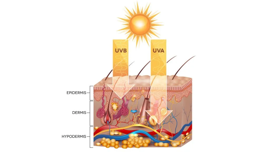
Ultraviolet (UV) radiation—both solar and artificial—stands out as the dominant external driver.  

   • UV-B (280–315 nm) directly induces cyclobutane pyrimidine dimers; if unrepaired, these mutations activate proto-oncogenes such as BRAF in melanocytes.  

   • UV-A (315–400 nm) penetrates deeper, generating reactive oxygen species that oxidise guanine bases and accelerate photo-ageing.  

   • Meta-analyses attribute 62–85 % of melanomas, basal-cell carcinomas (BCC) and cutaneous squamous-cell carcinomas (cSCC) to UV exposure, with intermittent blistering sunburn conferring the highest relative risk (RR 1.6–2.1) .

Artificial UV sources (tanning beds) amplify risk by 20–59 % for melanoma and 40–90 % for BCC/cSCC, especially when first exposure occurs before age 20.

Beyond light, immunosuppression (organ transplantation, biologic therapy, HIV) and inherited germline variants (CDKN2A, PTCH1) contribute.

Sun Riisk
Sun Risk

Who Is at the Highest Risk and Why?

Genetic and phenotypic traits  

• Fitzpatrick skin types I–III, red or blond hair, blue/green eyes and freckling all correlate with fewer epidermal melanin granules and therefore less natural UV shielding .  

• Family history of melanoma increases lifetime risk 2- to 4-fold .  

• Personal history of any skin cancer elevates the likelihood of subsequent lesions .

Environmental and lifestyle factors  

• Outdoor workers accumulate chronic UV dose, leading to cSCC on the head and neck (OR 2.2) .  

• Recreational sun-seekers often experience intermittent high-intensity exposure linked to superficial BCC and melanoma on the trunk .  

• Alcohol intake (>15 g/day) and ionising radiation (childhood radiotherapy) further increase BCC risk (OR 1.4 and 6.3, respectively) .

cancer risk.jpg
Cancer Risk

How Can You Spot Skin Cancer Early?

Self-examination remains the first line of defense. The widely validated ABCDE mnemonic—Asymmetry, Border irregularity, Colour variegation, Diameter >6 mm, Evolving—detects melanomas with 92 % sensitivity when two or more criteria are present .

A – Asymmetry  

   Draw an imaginary line through the lesion; halves should match.

B – Border  

   Scalloped, notched or poorly defined edges raise suspicion.

C – Colour  

   Look for multiple shades of brown, black, red, white or blue within the same mole.

D – Diameter  

   >6 mm is the classic threshold, though early melanomas can be smaller.

E – Evolving  

   Any change in size, shape, colour, elevation, or new symptom (bleeding, itching) warrants prompt evaluation.

ABCDEs of Skin Cancer

Non-melanoma clues  

• BCC: pearly papule with rolled border, telangiectasia or non-healing ulcer.  

• cSCC: hyperkeratotic nodule on sun-damaged skin or rapidly enlarging horn.

When Should You See a Doctor?

Immediate referral is indicated for any lesion that meets ABCDE criteria, persists beyond 4–6 weeks, ulcerates, or recurs after apparent healing . Additional red flags include:  

• New pigmented lesion in an adult >40 years.  

• Subungual or acral pigmentation (palms, soles) in darker skin phototypes.  

• Rapid growth (>25 % in 3 months).

Can an IBOOLO dermatoscope be used to detect skin cancer?

In July 2025, IBOOLO launched its newest pocket dermatoscope—the DE-500. Compared with the DE-400, the DE-500 adds a UV light mode and three-step brightness control. Most importantly, the mounting system has been completely re-engineered. While the DE-300 and DE-400 rely on a threaded back that must be screwed into the universal phone clip, the DE-500 features a circular magnetic ring. Simply align the dermatoscope with the magnetic clip included in the kit and it snaps securely into place—faster and more convenient than ever.

Whether you are a dermatologist in clinic or a concerned patient at home, the IBOOLO DE-500 equips you with clinical-grade optics, multi-modal lighting, and instant smartphone integration to detect melanoma, basal-cell carcinoma, and squamous-cell carcinoma earlier, more accurately, and with zero invasion.

IBOOLO DE-500 Dermatoscope

How Does a Dermatoscope Help Non-Invasively?

Traditional inspection has limitations: only 30–60 % of melanomas are correctly identified by naked-eye examination . A dermatoscope (e.g., IBOOLO’s pocket-size DE-500 or flagship DE-4100) bridges this gap by magnifying subsurface structures 10X under polarised and non-polarised light.

Key advantages  

Non-invasive: No biopsies or dyes; gel coupling suffices.  

Higher accuracy: Meta-analysis shows sensitivity rises to 93 % and specificity to 91 % when experienced clinicians use dermoscopy .  

Instant triage: Specific patterns—reticular network, globules, blue-white veil, leaf-like areas—differentiate melanoma from benign nevus or seborrheic keratosis.  

Patient empowerment: Handheld models that magnetically attach to a smartphone enable at-home documentation and tele-consultation, reducing unnecessary clinic visits.

Share this article

0

No products in the cart.

Ask

Have questions on gear or your order?

Our Gear Guides are here to help! Get personal advice from pro creatives

Name
Subject
Email address
How can we help?
Answers

Instant Answers

How can dermoscopy images be captured?

Dermoscopy images can be captured and stored in different ways, such as: • Using a smartphone or tablet with dermoscopic adapter, which consisted in the package.• Using a digital camera

Dermoscopy images can be captured and stored in different ways, such as:

• Using a smartphone or tablet with dermoscopic adapter, which consisted in the package.
• Using a digital camera with dermoscopic adapter, there’s 49mm screw size camera adapter available to order now.

Compatible phone/tablet models:
All iPhone models, 95% Android phones, 90% tablet. For phone/tablet size in 5.25-14mm

Compatible camera models:
All camera with built 49mm filter screw, such as Canon EOS 70D, 80D, 90D; Canon EOS R7, R10, R50, R100; Canon M100, M200, M50, Mark II; Canon G7X Mark III, Sony ZV-1

How can I connect my phone to my dermatoscope?

There’s universal phone adapter for all our dermoscopes. Please check the installation procedure bellow or watch operation guide. Smartphone Connector (1) Place phone adapter screw in the center of smartphone’s

There’s universal phone adapter for all our dermoscopes. Please check the installation procedure bellow or watch operation guide.

Smartphone Connector

(1) Place phone adapter screw in the center of smartphone’s main camera.
(2) Screw magnet attachment on phone adapter.
(3) Put dermoscope’s back ring and magnet attachment together

Take The Best Images

You need to adjust the focus ring after the dermoscpe connected on smartphone to get the best images.

How can I clean my dermoscopy after usage?

Cleaning your dermoscopy after usage is important to prevent cross-contamination and infection. The cleaning method may vary depending on the type and model of your dermoscopy, so you should always

Cleaning your dermoscopy after usage is important to prevent cross-contamination and infection. The cleaning method may vary depending on the type and model of your dermoscopy, so you should always follow the manufacturer’s instructions. However, some general steps are:

• Turn off and disconnect your dermoscopy from any power source or device.

• Wipe off any visible dirt or debris from the dermoscopy with a soft cloth or tissue.

• Disinfect the dermoscopy with an alcohol-based wipe or spray, or a disinfectant solution recommended by the manufacturer. Make sure to cover all surfaces, especially the lens and contact plate.

• Let the dermoscopy air dry completely before storing it in a clean and dry place.

• Do not use abrasive or corrosive cleaners, solvents, or detergents that may damage the dermoscopy.

• Do not immerse the dermoscopy in water or any liquid, unless it is waterproof and designed for immersion.

You should clean your dermoscopy after each use, or at least once a day if you use it frequently. You should also check your dermoscopy regularly for any signs of damage or malfunction, and contact the manufacturer or service provider if needed.

Polarized VS Non-polarized Dermoscopy

A dermoscopy is a device that allows the examination of skin lesions with magnificationand illumination. By revealing subsurface structures and patterns that are not visible tothe naked eye. It can

A dermoscopy is a device that allows the examination of skin lesions with magnificationand illumination. By revealing subsurface structures and patterns that are not visible tothe naked eye. It can improve the diagnose accuracy of skin lesions, such as melanoma,basal cell carcinoma, seborrheic keratosis, etc.

There are two main types of dermoscopy: Non polarized and polarized dermoscopy.We’ve fitted most of our dermoscopys with polarized and non-polarized light. They canbe used in multiple skin structures.

Non-polarized contact Mode

In non-polarized mode, the instrument can provide information about the superficialskin structures, such as milia-like cysts, comedo-like openings, and pigment in theepidemis.

The dermoscopy requires applying a liquid such as mineral oil or alcohol to the skin andplacing the lens in contact with the skin. This reduces surface reflection and enhancesthe view of subsurface structures.

Image with non-polarized light (DE-3100)

Polarized contact Mode

In polarized mode, the instrument allows for visualization for deeper skin structures,such as blood vessels, collagen, and pigment in the dermis.

The dermoscopy does not need to be in contact with the skin or use any liquid. Theirpolarized light can help to eliminate surface reflection and allow visualization ofvascular structures.

Image with polarized light (DE-3100)

Polarized non-contact Mode

The dermoscopy can also use polarized light to examine the skin without direct contact.

In polarized non-contact mode, the instrument allows for examination infected areasand lesions that are painful for the patient, or the difficult to contact pigmented lesions,such as nails and narrow areas.

The contact plate should be removed in this mode, and it does not require applying aliquid to the skin. As it doesn’t require pressure or fluid application on the skin, it canalso avoid cross-contamination and infection risk.

Image in polarized non-contact mode (DE-3100)

How effectiveness is dermoscopy

Compared with visual inspection, the dermoscopy can be used to capture and store skin lesion photos, which play an important role in early skin cancer examination. The dermoscopy allows the

Compared with visual inspection, the dermoscopy can be used to capture and store skin lesion photos, which play an important role in early skin cancer examination.

The dermoscopy allows the examination of skin lesions with magnification and illumination. This can be greatly avoiding the factors that cause interference to visual detection. Such as lighting, skin color, hair and cosmetics.

Several studies have demonstrated that dermoscopy is useful in the identification of melanoma, when used by a trained professional.

It may improve the accuracy of clinical diagnosis by up to 35%
It may reduce the number of harmless lesions that are removed
In primary care, it may increase the referral of more worrisome lesions and reduce the referral of more trivial ones

A 2018 Cochrane meta-analysis published the accuracy of dermoscopy in the detection.

Table 1. Accuracy of dermoscopy in the detection of melanoma in adults
Detection Method Sensitivity, % Specificity, % Positive Likelihood Ratio NegativeLikelihood Ratio
Visual inspection alone (in person) 76 75 3.04 0.32
Dermoscopy with visual inspection (in person) 92 95 18 0.08
Image-based visual inspection alone (not in person) 47 42 0.81 1.3
Dermoscopy with image-based visual inspection (not in person) 81 82 4.5 0.23
ROC—receiver operating characteristic. *Estimated sensitivity calculated on the summary ROC curve at a fixed specificity of 80%.

As we can see, the dermoscope can improve the accuracy of diagnosis of skin lesions, especially melanoma.

Table 1. Accuracy of dermoscopy in the detection of melanoma in adults
Detection Method Sensitivity, % Specificity, % Positive Likelihood Ratio NegativeLikelihood Ratio
Visual inspection alone (in person) 79 77 3.4 0.27
Dermoscopy with visual inspection (in person) 93 99 93 0.07
Image-based visual inspection alone (not in person) 85 87 6.5 0.17
Dermoscopy with image-based visual inspection (not in person) 93 96 23 0.07
ROC—receiver operating characteristic. *Estimated sensitivity calculated on the summary ROC curve at a fixed specificity of 80%.

Characteristics of the dermatoscopic structure of the skin lesions include:

• Symmetry or asymmetry
• Homogeny/uniformity (sameness) or heterogeny (structural differences across the lesion)
• Distribution of pigment: brown lines, dots, clods and structureless areas
• Skin surface keratin: small white cysts, crypts, fissures
• Vascular morphology and pattern: regular or irregular
• Border of the lesion: fading, sharply cut off or radial streaks
• Presence of ulceration

There are specific dermoscopic patterns that aid in the diagnosis of the following pigmented skin lesions:

• Melanoma
• Moles (benign melanocytic naevus)
• Freckles (lentigos)
• Atypical naevi
• Blue naevi
• Seborrhoeic keratosis
• Pigmented basal cell carcinoma
• Haemangioma

We use cookies on this website to provide a better user experience. By continuing to browse the website, you are giving your consent to receive cookies on this site. For more details please read our Privacy Policy.

Hot Search Terms