


Article
Dermatoscope: The Third Eye of Skin Doctors. Then What is A Dermatoscope and Dermoscopy Meaning?
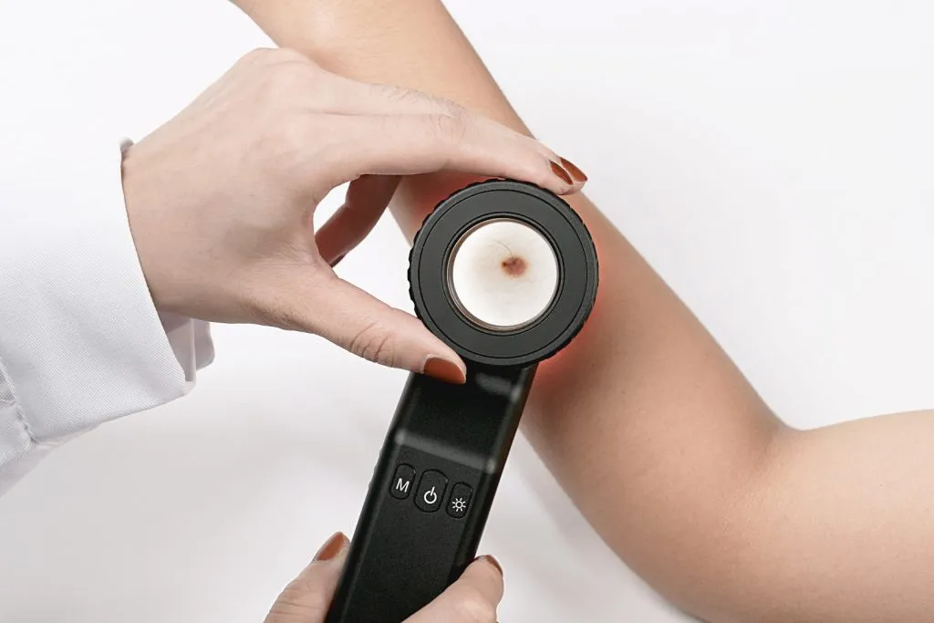
What is a dermatoscope ? Dermatoscope (Dermoscopy) is a handheld optical device usually used to examine skin or hair much more accurately. It combines high quality magnify lens and powerful lighting system to enhance the view of deeper skin. Without any side effective or adverse reactions, also avoiding unnecessary biopsies and surgeries, it is very…
what is a dermatoscope | dermoscopy meaning - IBOOLO
IBOOLO Discover the power of the dermatoscope and uncover the true meaning of dermoscopy. Explore this advanced diagnostic tool, capabilities that surpass the naked eye or a simple magnifying glass.
What is a Dermatoscope? Understanding Dermoscopy Meaning and Applications
What is a dermatoscope?
What exactly is a dermatoscope, and what is the meaning of dermoscopy in modern dermatology practice? A dermatoscope is a hand-held visual aid device used by doctors and healthcare professionals to examine and diagnose skin lesions and diseases. This non-invasive tool is particularly valuable in diagnosing conditions like melanoma, while also enabling detailed examination of the scalp, hair, and nails. Through its specialized magnifying lens and illumination system, it allows users to observe microscopic skin structures that are invisible to the naked eye.
Key features of a dermatoscope include:
- Magnification (typically 10x to 20x)
- Built-in light source (LED or halogen)
- Polarized or non-polarized light options
- Contact or non-contact examination capabilities
What does dermoscopy meaning?
Dermoscopy (also known as dermatoscopy or chemiluminescence microscopy) is the examination technique performed using a dermatoscope. This diagnostic method allows healthcare providers to observe skin structures and patterns that are not visible to the naked eye. Through its specialized magnifying lens and illumination system, dermoscopy enhances the visualization of deeper skin layers, making it an essential procedure in modern dermatological practice.
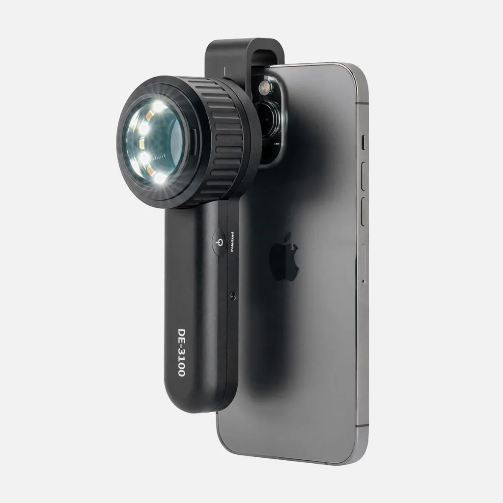
How Does a Dermatoscope Work?
A dermatoscope works by combining several key components: A powerful LED light source that illuminates the skin, Magnifying lens (typically 10x magnification), Cross-polarized filters that reduce surface reflection and A contact plate that flattens the skin surface for better viewing. The dermatoscope eliminates surface glare and allows visualization of deeper skin structures by using either contact (with immersion fluid) or non-contact polarized light techniques.
What Happens During a Dermatoscopy?
During a dermoscopy examination:
1. The doctor cleans the area to be examined
2. If using contact dermoscopy, they apply a small amount of immersion fluid (oil, alcohol, or gel)
3. The dermatoscope is placed against the skin
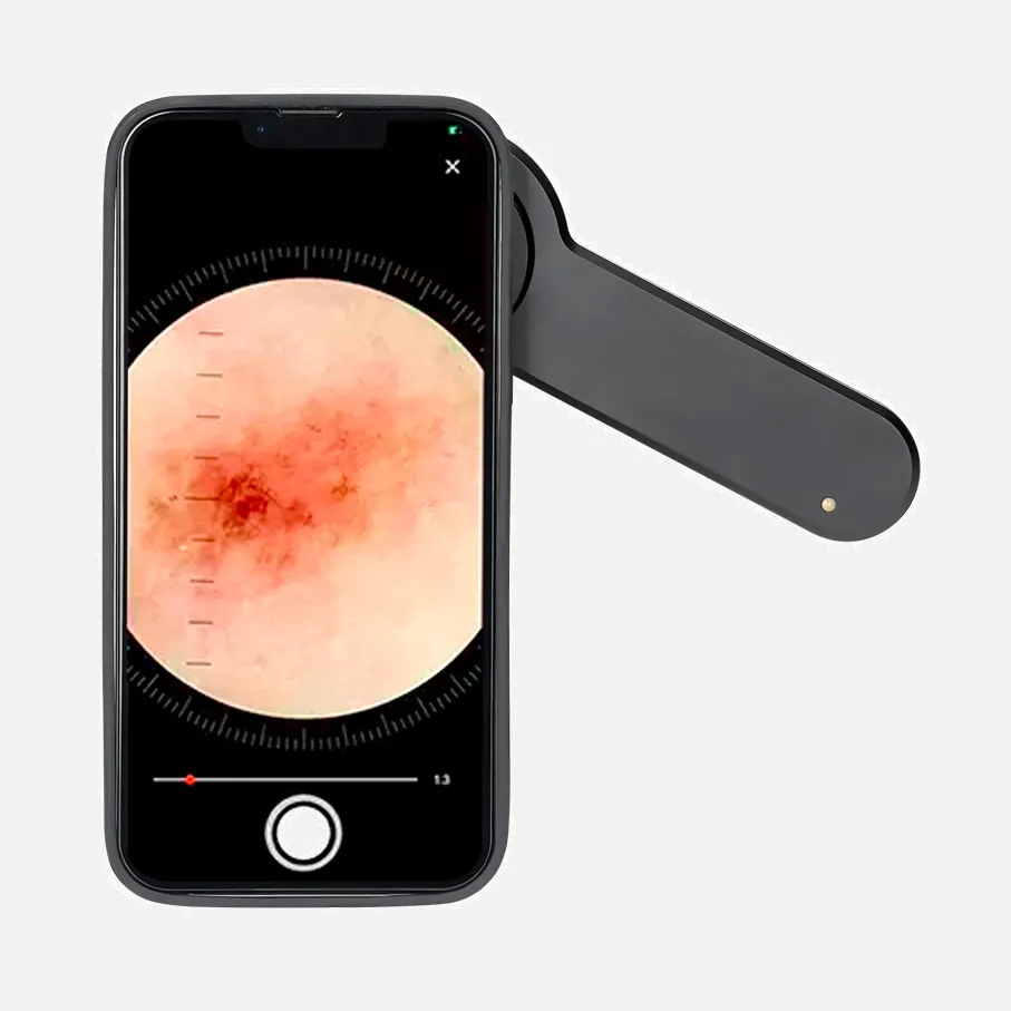
4. The physician examines the lesion's patterns, colours, and structures
5. Images may be captured for documentation
6. Multiple lesions can be examined in a single session
What Can a Dermatoscope See?
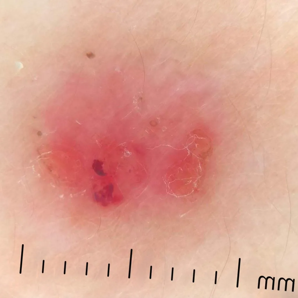
A dermatoscope can visualize: Pigment network patterns, Blood vessel structures, Color variations within lesions, Structural features not visible to the naked eye, Early signs of skin cancer, Different types of skin growths, Hair follicle abnormalities, Mite infestations (like scabies).
Are Dermatoscopes Accurate?
Dermoscopy significantly improves diagnostic accuracy compared to the naked eye examination. Studies show that using a dermatoscope increases melanoma detection accuracy by 10-27% compared to visual inspection alone. However, accuracy depends on several factors.
Factors Affecting Accuracy
1. Operator Experience: Training level of the practitioner, Familiarity with dermoscopic patterns, Regular practice and usage.
2. Technical Factors: Quality of the dermatoscope, Proper lighting conditions, Correct use of immersion fluid when required, and Image resolution (for digital dermatoscopes).
3. Patient Factors: Skin condition (presence of blood, scabs, or scales), Location of the lesion, Patient movement during the examination, Skin type and pigmentation.
Different Types of Dermatoscopes
1. Traditional Contact Dermatoscopes: Require immersion fluid, Direct skin contact, Typically non-digital, Usually more affordable.
2. Polarized Light Dermatoscopes: No need for immersion fluid, Can be used with or without contact, Better visualization of vascular structures, Reduced glare.
3. Digital Dermatoscopes: Capture and store images, Allow for lesion monitoring over time, Can integrate with electronic medical records, Enable telemedicine consultations and Higher costs but more features.
4. Hybrid Dermatoscopes: Combine both polarized and non-polarized viewing, Offer both contact and non-contact options, Versatile for different examination needs and Popular among experienced practitioners.
The field of dermoscopy continues to evolve with new technological advances, making dermatoscopes increasingly essential tools in skin examination and cancer detection. Whether using a basic contact dermatoscope or an advanced digital system, these devices significantly enhance a practitioner's ability to evaluate skin conditions accurately and early.
Limitations and Considerations
While understanding what a dermatoscope is and the meaning of dermoscopy is crucial, it's also important to recognize its limitations:
1. Learning Curve: Proper interpretation of dermoscopic images requires training and experience.
2. Not a Standalone Diagnostic Tool: This should be used in conjunction with clinical examination and patient history.
3. Quality Variability: The effectiveness can vary based on the quality of the dermatoscope and the skill of the user.
Future of Dermoscopy
The meaning of dermoscopy continues to evolve with technological advancements:
1. Artificial Intelligence Integration: AI algorithms are being developed to assist in lesion analysis.
2. Teledermoscopy: Remote consultations using dermoscopic images are becoming more common.
3. 3D Imaging: Advanced systems are incorporating three-dimensional mapping of skin lesions.
4. Multispectral Imaging: Emerging technologies allow the examination of skin under different light wavelengths.
Understanding what a dermatoscope is and the full meaning of dermoscopy is essential for both dermatology professionals and patients. As a powerful diagnostic tool, the dermatoscope has revolutionized skin examination, offering a bridge between clinical observation and histological analysis. Its ability to provide detailed, non-invasive skin assessment has made it an indispensable instrument in modern dermatology practice, significantly improving the accuracy of skin cancer detection and the management of various skin conditions.
Recommended reading
Download MSDS Report – IBOOLO
Shenzhen Iboolo Optics Co.Ltd founded in 2012,is located in the beautiful city of Shenzhen, Guangdong. Our main products including Woods Lamp, Dermatoscope, Microscope and Macro lens, etc.
basal cell carcinoma dermoscopy – IBOOLO
Shenzhen Iboolo Optics Co.Ltd has been specialized in researching and manufacturing industrial Dermatoscope, Microscope, Macro lens and Woods Lamp, since 2012. As a professional camera lens supplier, we have excellent teams who focus on products development & design, quality control & inspection and company running.
FAQs – IBOOLO
Compared with visual inspection, the dermoscopy can be used to capture and store skin lesion photos, which play an important role in early skin cancer examination. The dermoscopy allows the examination of skin lesions with magnification and illumination. This can be greatly avoiding the factors that cause interference to visual detection. Such as lighting, s...
What is a dermatoscope ?
Dermatoscope (Dermoscopy) is a handheld optical device usually used to examine skin or hair much more accurately. It combines high quality magnify lens and powerful lighting system to enhance the view of deeper skin. Without any side effective or adverse reactions, also avoiding unnecessary biopsies and surgeries, it is very helpful for doctors to diagnose skin lesions,such as infection skin disease,pigmented skin disease,inflammatory skin disease,vascular skin disease, onychosis and so on.Only by clearly understanding dermoscopy meaning,people can use it in high efficiency.
Types of dermatoscope
There are three main types of dermatoscopy, polarized dermatoscopy ,nonpolarised dermatoscope and amber dermatoscopy.
•Polarized dermatoscopy: To eliminate surface glare and reflection of the skin by utilizing polarized light, it gets deeper peer of skin and can clearly display the dermis structure.No need to use liquid on skin surface and no need to touch skin,it is more safety for diagnosing skin lesion and avoiding the risk of cross infection.
•Nonpolarized dermatoscopy: Nonpolarized dermoscopy can clearly show the cuticular layer of skin with or without liquid medium.
• Amber dermatoscopy: By using amber dermoscopy, we can clearly inspect the outline of structure of skin dermis and epidermis from its shape,size,color,bugle,etc.
As we can know from above, three types of dermoscopy provide complementary skin information, so that diagnose will be more accurte and comprehensive.
Principle of dermatoscope
Dermatoscopy combines the medical technology and principle of physics and optics. Dermatologists can observe skin lesions accurately and objectively by dermatoscopy, seeing through appearance to the essence, seeing through surface layer to deep layer,seeing through epidermal layer to dermal layer,seeing through naked eye to lens.
For the observation of skin lesion,dermatoscopy plays a role far more than a magnifier. It uses polarizing filter to filter out the diffuse reflection light and then with the aid of optical,
it filters out the diffuse light of the epidermis by using polarizing filters, and then through the optical amplification equipment to observe the skin including under the epidermis, dermal-epidermis junction, pigment of the dermal papillary layer and blood vessels such skin structures which cannot be saw by naked eyes. Knowing dermoscopy meaning deeply will help skin doctors work more easily and confidently.
What is the use of dermastocope?
By using dermatoscopy, dermatologists can diagnose skin lesions and diseases more accurately. And dermascope is easy to operate. The whole process of using dermoscope is simple and painless. With the help of dermoscopy, person can examine their own hair,scalp,skin, and nails clearly. Then they can send images captured by phone or tables under dermoscope to dermatologists for analysis.
Then what is a dermatoscope? Dermatscopy is hand-held device which is also known as dermoscope. It usually used in skin lesions examination by dermatologists. It reveals structures of skin surface and subsurface invisible to naked eyes by the technology combining optics and physics. Dermatscope creates a process of green,painless and noninvasive. That is the dermoscopy meaning. There are common uses of dermatoscope as below:
Common Uses:
1, Detecting hair loss and alopecia areata
2, Distinguishing melanoma from pigmented naevus
3, Identifying skin cancer from its benign lesion
4, Inspecting other skin disease such as lichen planus, vitiligo and scabies
Which kind of diseases can be detected by dermoscopy?
By dermoscopy, doctors can detecte skin lesions, skin cancer, dermatitis, infections, acne, hair loss and nail problmes, etc.
Common diseases:
Begin melanoma, early skin tumor, basal cell carcinoma, benign and malignant cascular lesions, etc.
Many skin diseases can be examined under dermoscopy otherwise routine naked eyes cannot pick them out. That is dermoscopy meaning to the whole medical world.
What will effect the examination of dermatoscopy?
What is a dermatoscope? Dermoscopy is a device usually used by skin doctors for examining skin lesions. It is hand-held and easy to operate. It can greatly enhance the view of inspection to improve the diagnostic accuracy. Otherwise, there are some factors which will effect the results of dermoscopy examination, such as lighting conditionS, ointment on skin surface, device quality and setting,etc.
1.Lighting conditions
Poor or Hard light both will affect dermatoscope examination. So proper light is very necessary for the accurate examination of dermatoscope.
2.Ointment on skin surface
Ointment will bring disturb for dermatoscope examination. Before dermatoscopy examination, people do not apply any ointment to the skin to avoild misdiagnosis. It is better to keep the skin clear and try as far as possible.
3.Device quality and setting
Good quality for dermatoscopy is crucial for dermatologists inspection. And its setting whether can be adjustable matters the possibility of customization for different kinds of skin lesions.
Can dermatoscope detect skin cancer?
Yes, dermatoscope can detect skin cancer. As we know dermoscopy meaning for dermatologists, with magnification and illumination of dermatscope, examination by dermatoscope increases not only specificity but also sensitivity for skin cancer. It can detect smaller and thinner skin cancer from its structure,pattern,shape which may be missed by naked eyes, increasing biopsy of melanomas,reducing the the biopsy of lesion beginning.
What does skin cancer look like under dermatoscope?
When examining skin cancer under a dematoscope, there are various features presented with valuable information which invisible by routine inspection.Then what is dermatoscope? Dermatoscope is often also called epiluminescence microscopy, which is a hand-held aid device equip skin doctors observing skin cancer more effectively. Because dermatoscope enhance the visualization of skin lesions. There are types of skin cancer with diverse key features showed under dermatscope, such as multiple brown dots, blue-white veil,scar-like depigmentation, pseudopods, squamous cell carcinoma, radial streamlines,peripheral black spots/globules, multiple colors, broad nedwork, focal sharp cut odd boundary, malignant melanoma, basel cell carcinoma ,ect.
Is dermatoscope accurate?
People will wonder is dermoscope accurate, so first let us to understand what is a dermatoscope?
Dermatoscopy, also known as a dermoscope, is a dependable optical tool for detecting skin disease or skin problems. It enhances the visibility by polarized light or non-polarized light or amber light combining magnification. Particularly in experienced hands, it can detect and diagnose skin situations much more clearly and accurately.
Compared with routine examination by naked eyes, dermatologist can use dermatoscope uniting phone or tablet to capture photos of skin lesion layers from epidermis to dermis. It not only saves time for people in inspection but also creates much more convenience for analysis.
Report from Oncology Section of the Skin and Cancer Unit of NYU Langone Medical Center, that dermatologist can only diagnose 65%~80% of melanomas. Accuracy of diagnosis by dermatologists is only 64% (Grin et al.,1990). But by using dermoscopy, it increases the accuracy of diagnosis by 10~27% (Kittler et al.,2002)
Dermoscopy meaning is very important for various of skin examinations, especially in uncommon skin problems. Even though, it still needs to combine the clinical knowledge and clinical experience to diagnose skin lesions much more accuracy.


