


Article
Dermatoscope: The Third Eye of Skin Doctors. Then What is A Dermatoscope and Dermoscopy Meaning?
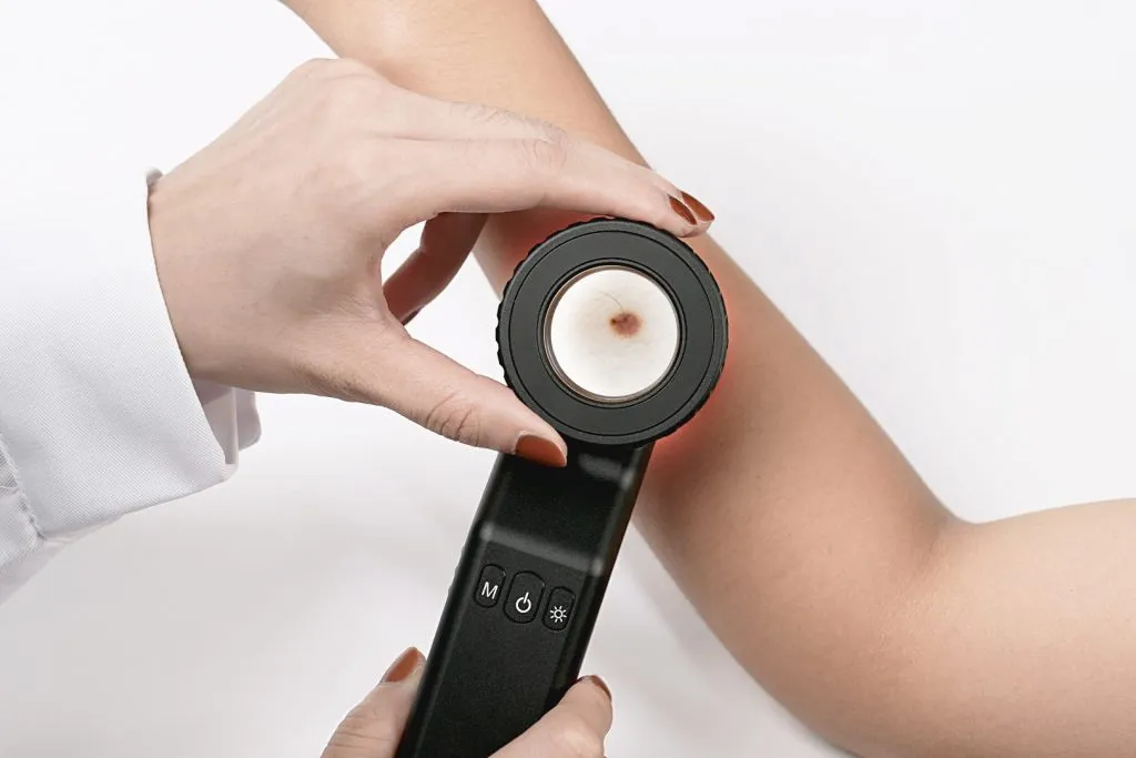
What is a dermatoscope ? Dermatoscope (Dermoscopy) is a handheld optical device usually used to examine skin or hair much more accurately. It combines high quality magnify lens and powerful lighting system to enhance the view of deeper skin. Without any side effective or adverse reactions, also avoiding unnecessary biopsies and surgeries, it is very…
Dermoscopy Meaning & Dermoscopi Guide: A Comprehensive Overview | IBOOLO
Understand the true dermoscopy meaning and what a Dermoscopi is. IBOOLO expert guide breaks down how this tool works, its applications in diagnosing skin conditions, and why it's a game-changer for skin health.
The Ultimate Guide to Dermoscopy: Understanding the Dermoscopy Meaning and the Role of the Dermoscopi
In modern dermatology, few tools are as crucial as the Dermoscopi. This handheld device has revolutionized the way doctors examine and diagnose skin conditions, offering insights that are simply impossible to see with the naked eye. But what exactly is a Dermoscopi, and what is the true dermoscopy meaning? This comprehensive guide will take you on a deep dive into the world of dermoscopy, exploring its history, technology, and why it has become an indispensable part of skin cancer diagnosis and beyond.
What is a Dermoscopi? A Professional Definition
A Dermoscopi is a specialized, non-invasive diagnostic tool used by medical professionals to examine skin lesions, hair, and nails. By combining a powerful magnification lens (typically 10x to 20x) with a high-quality illumination system, the Dermoscopi allows for a detailed, magnified view of subsurface skin structures and patterns. This process, known as dermoscopy, bridges the gap between a clinical visual inspection and a microscopic pathological analysis, significantly improving diagnostic accuracy.
Key Components of a Modern Dermoscopi:
- Magnifying Lens: Provides a magnified, clear image of the lesion.
- Illumination System: A high-intensity light source (usually LED) that brightly illuminates the skin surface.
- Polarized and Non-Polarized Filters: Advanced models feature filters that can be toggled to eliminate surface glare and reveal deeper structures.
- Contact Plate: A clear plate that touches the skin, often used with a liquid medium to improve light transmission.
Decoding the Dermoscopy Meaning: A Deeper Look
The term dermoscopy comes from the Greek words "derma" (skin) and "skopein" (to view). Fundamentally, the dermoscopy meaning is "the visual examination of the skin." However, in modern medical practice, it signifies a specific, highly technical procedure that utilizes a Dermoscopi to observe a multitude of intricate features. These features, often referred to as "dermoscopic patterns," include the morphology of blood vessels, the organization of pigment networks, and the presence of specific structural features like milia-like cysts or comedo-like openings. By interpreting these patterns, a dermatologist can more accurately differentiate between a harmless mole and a potentially life-threatening skin cancer like melanoma or basal cell carcinoma.
The History and Evolution of the Dermoscopi
While the concept of dermoscopy dates back to the late 19th century, the first practical Dermoscopi was developed in the 1920s. Early devices were simple light microscopes that required a layer of oil on the skin to reduce surface reflection. The true revolution in dermoscopy came with the introduction of cross-polarization technology in the 1990s. This innovation allowed for glare-free, non-contact examination of the skin, making the process faster, more comfortable for the patient, and more revealing for the practitioner. Today's digital Dermoscopi takes this a step further, allowing for image capture, storage, and long-term lesion monitoring, which is a cornerstone of modern skin cancer surveillance.
How Does a Dermoscopi Work? The Science of Visualization
A Dermoscopi works by using a combination of magnification and a controlled light source to eliminate surface interference and provide a clear view of subsurface structures. The two primary modes of operation are:
1. Non-Polarized (Contact) Dermoscopy
This traditional method requires direct contact with the skin. A small amount of an immersion fluid, such as mineral oil or alcohol, is applied to the lesion. This fluid acts as a bridge, reducing light scattering at the skin's surface and allowing light to penetrate deeper. This mode is particularly useful for visualizing superficial epidermal features, like those found in seborrheic keratosis or pigmented spots.
2. Polarized (Non-Contact) Dermoscopy
Polarized dermoscopy uses a special filter system to block surface-reflected light. This allows the practitioner to see directly into the dermis without any need for contact or immersion fluid. This mode is a game-changer for visualizing vascular structures (blood vessels) and deeper dermal pigment, which are crucial indicators of malignancy. The ability to perform a non-contact exam is also invaluable for painful or infected lesions, or for areas that are difficult to access.
What Can a Dermoscopi See? A Comprehensive List of Structures
By using a Dermoscopi, a trained professional can identify and analyze a wide range of features, helping to inform a more accurate diagnosis. The structures and patterns visible with a Dermoscopi include:
- Pigment Network: The most important feature for diagnosing pigmented lesions, revealing patterns that can be regular (benign) or irregular (malignant).
- Globules and Clods: Spherical or oval structures that represent nests of melanocytes.
- Streaks and Pseudopods: Linear extensions at the border of a lesion, often associated with melanoma.
- Vascular Patterns: The arrangement and shape of blood vessels, which can be highly indicative of different skin tumors.
- Blue-white veil: A bluish-gray haze that is a hallmark feature of melanoma.
- Milia-like cysts and Comedo-like openings: Key features of seborrheic keratosis.
The Clinical Value of the Dermoscopi: Why Dermoscopy Matters
The dermoscopy meaning in a clinical setting is directly tied to its ability to improve diagnostic accuracy. Studies have shown that when a Dermoscopi is used by a trained professional, the accuracy of melanoma detection can increase by up to 27% compared to examination with the naked eye alone. This leads to earlier detection, better patient outcomes, and a reduction in unnecessary biopsies of benign lesions.
The Dermoscopi is not just for pigmented lesions. It is also a powerful tool for diagnosing a wide range of other skin conditions, including:
- Psoriasis and other inflammatory skin diseases.
- Fungal infections and scabies.
- Hair and scalp disorders (trichoscopy).
- Nail abnormalities (onychoscopy).
Factors Influencing the Accuracy of a Dermoscopy Examination
While the Dermoscopi is a highly effective tool, its accuracy depends on several key factors:
1. Operator Expertise
The interpretation of dermoscopic patterns requires extensive training and experience. A skilled practitioner can identify subtle features that may be missed by a novice. This is why continuous education and practice are essential for any medical professional using a Dermoscopi.
2. Quality of the Dermoscopi
The optical and lighting quality of the Dermoscopi directly impacts the clarity of the image. A high-resolution Dermoscopi with superior polarization capabilities will provide a more detailed and accurate view, making diagnosis easier and more reliable. This is where investing in a professional-grade device is crucial.
The Future of Dermoscopy: Merging Technology and Medical Practice
The dermoscopy meaning continues to expand as technology evolves. The future of the Dermoscopi lies in its integration with advanced digital systems:
- AI-Assisted Diagnostics: Artificial intelligence algorithms are being trained on vast databases of dermoscopic images to provide instant, data-driven analysis and a second opinion.
- Teledermoscopy: Digital Dermoscopi images can be securely transmitted to specialists for remote consultation, bridging geographical gaps in healthcare.
- Multispectral Imaging: Emerging devices are using different wavelengths of light to reveal new information about skin composition and blood flow.
The Dermoscopi is More Than Just a Magnifying Glass
Understanding what a Dermoscopi is and the full dermoscopy meaning is essential for anyone in the field of dermatology. It is not just a simple visual aid; it is a sophisticated diagnostic instrument that has fundamentally changed how skin conditions are evaluated and treated. Its ability to provide a non-invasive, high-resolution view of skin structures has made it an indispensable instrument in the fight against skin cancer and a vital tool for managing a wide array of dermatological conditions. As a cornerstone of modern skin examination, the Dermoscopi will continue to play a pivotal role in the future of medical diagnostics, providing a bridge between clinical observation and scientific precision.
Recommended reading
High Quality Dermoscopy Meaning Created in Our Products Supply Based in China - IBOOLO
Our China products supply hub couples world-class portability with elite precision, using seasoned expertise to develop high quality dermoscopy meaning for flawless skin visualization anywhere through compact size.
China Skin Cancer Dermoscopy Products Supply Specializes in Professional Items - IBOOLO
Our China products supply creates clinical quality Professional skin cancer dermoscopys enabling powerful skin magnification from anywhere through thoughtful craftsmanship.
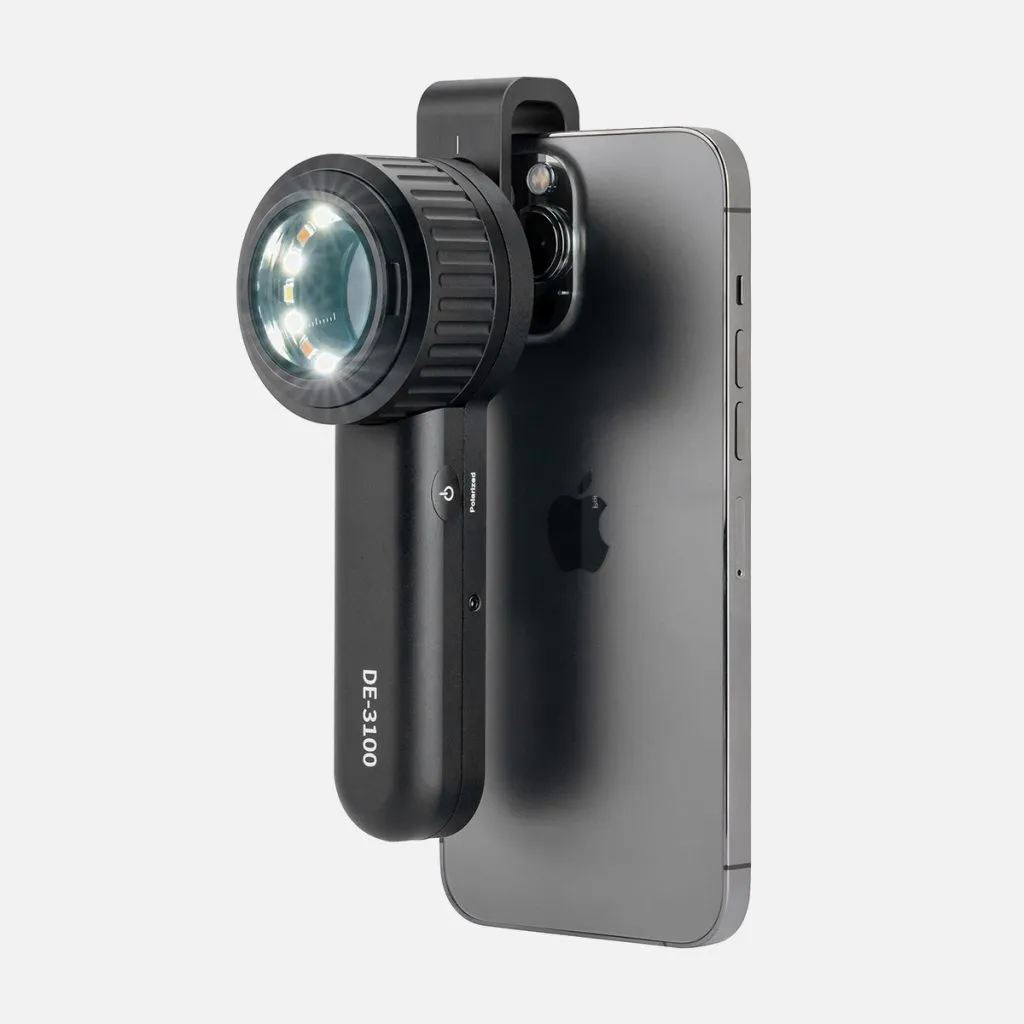
China Products Supply Provides Wholesale Dermatoscope Phone Attachments for Clients - IBOOLO
As an expert China products supply, we use exacting wholesale production methods to manufacture high-quality dermatoscope phone attachment solutions tailored for every customer.
What is a dermatoscope ?
Dermatoscope (Dermoscopy) is a handheld optical device usually used to examine skin or hair much more accurately. It combines high quality magnify lens and powerful lighting system to enhance the view of deeper skin. Without any side effective or adverse reactions, also avoiding unnecessary biopsies and surgeries, it is very helpful for doctors to diagnose skin lesions,such as infection skin disease,pigmented skin disease,inflammatory skin disease,vascular skin disease, onychosis and so on.Only by clearly understanding dermoscopy meaning,people can use it in high efficiency.
Types of dermatoscope
There are three main types of dermatoscopy, polarized dermatoscopy ,nonpolarised dermatoscope and amber dermatoscopy.
•Polarized dermatoscopy: To eliminate surface glare and reflection of the skin by utilizing polarized light, it gets deeper peer of skin and can clearly display the dermis structure.No need to use liquid on skin surface and no need to touch skin,it is more safety for diagnosing skin lesion and avoiding the risk of cross infection.
•Nonpolarized dermatoscopy: Nonpolarized dermoscopy can clearly show the cuticular layer of skin with or without liquid medium.
• Amber dermatoscopy: By using amber dermoscopy, we can clearly inspect the outline of structure of skin dermis and epidermis from its shape,size,color,bugle,etc.
As we can know from above, three types of dermoscopy provide complementary skin information, so that diagnose will be more accurte and comprehensive.
Principle of dermatoscope
Dermatoscopy combines the medical technology and principle of physics and optics. Dermatologists can observe skin lesions accurately and objectively by dermatoscopy, seeing through appearance to the essence, seeing through surface layer to deep layer,seeing through epidermal layer to dermal layer,seeing through naked eye to lens.
For the observation of skin lesion,dermatoscopy plays a role far more than a magnifier. It uses polarizing filter to filter out the diffuse reflection light and then with the aid of optical,
it filters out the diffuse light of the epidermis by using polarizing filters, and then through the optical amplification equipment to observe the skin including under the epidermis, dermal-epidermis junction, pigment of the dermal papillary layer and blood vessels such skin structures which cannot be saw by naked eyes. Knowing dermoscopy meaning deeply will help skin doctors work more easily and confidently.
What is the use of dermastocope?
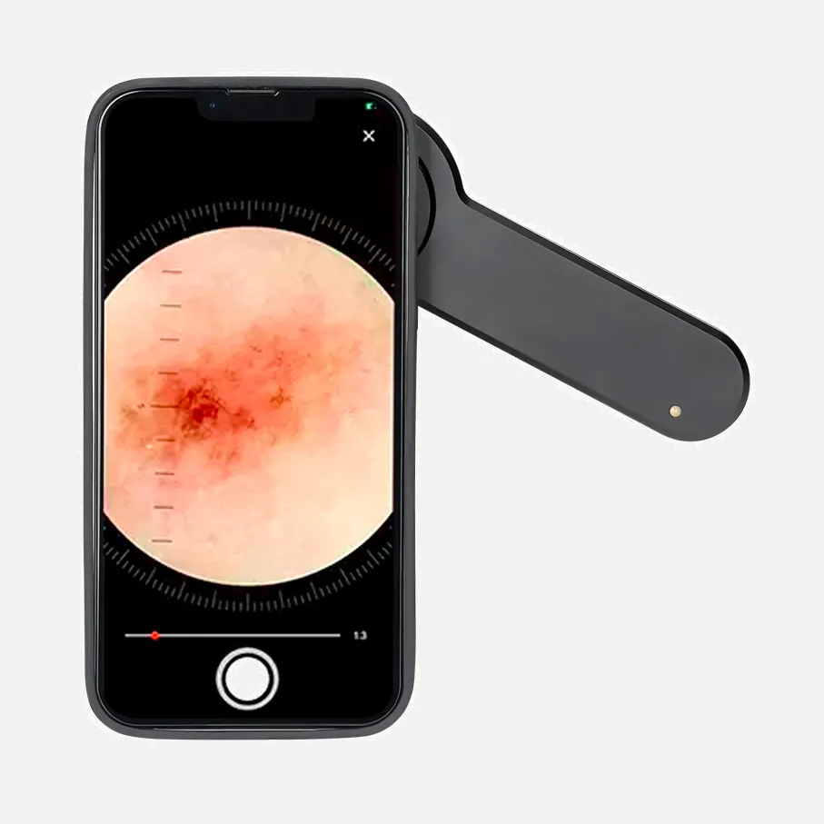
By using dermatoscopy, dermatologists can diagnose skin lesions and diseases more accurately. And dermascope is easy to operate. The whole process of using dermoscope is simple and painless. With the help of dermoscopy, person can examine their own hair,scalp,skin, and nails clearly. Then they can send images captured by phone or tables under dermoscope to dermatologists for analysis.
Then what is a dermatoscope? Dermatscopy is hand-held device which is also known as dermoscope. It usually used in skin lesions examination by dermatologists. It reveals structures of skin surface and subsurface invisible to naked eyes by the technology combining optics and physics. Dermatscope creates a process of green,painless and noninvasive. That is the dermoscopy meaning. There are common uses of dermatoscope as below:
Common Uses:
1, Detecting hair loss and alopecia areata
2, Distinguishing melanoma from pigmented naevus
3, Identifying skin cancer from its benign lesion
4, Inspecting other skin disease such as lichen planus, vitiligo and scabies
Which kind of diseases can be detected by dermoscopy?
By dermoscopy, doctors can detecte skin lesions, skin cancer, dermatitis, infections, acne, hair loss and nail problmes, etc.
Common diseases:
Begin melanoma, early skin tumor, basal cell carcinoma, benign and malignant cascular lesions, etc.
Many skin diseases can be examined under dermoscopy otherwise routine naked eyes cannot pick them out. That is dermoscopy meaning to the whole medical world.
What will effect the examination of dermatoscopy?
What is a dermatoscope? Dermoscopy is a device usually used by skin doctors for examining skin lesions. It is hand-held and easy to operate. It can greatly enhance the view of inspection to improve the diagnostic accuracy. Otherwise, there are some factors which will effect the results of dermoscopy examination, such as lighting conditionS, ointment on skin surface, device quality and setting,etc.
1.Lighting conditions
Poor or Hard light both will affect dermatoscope examination. So proper light is very necessary for the accurate examination of dermatoscope.
2.Ointment on skin surface
Ointment will bring disturb for dermatoscope examination. Before dermatoscopy examination, people do not apply any ointment to the skin to avoild misdiagnosis. It is better to keep the skin clear and try as far as possible.
3.Device quality and setting
Good quality for dermatoscopy is crucial for dermatologists inspection. And its setting whether can be adjustable matters the possibility of customization for different kinds of skin lesions.
Can dermatoscope detect skin cancer?
Yes, dermatoscope can detect skin cancer. As we know dermoscopy meaning for dermatologists, with magnification and illumination of dermatscope, examination by dermatoscope increases not only specificity but also sensitivity for skin cancer. It can detect smaller and thinner skin cancer from its structure,pattern,shape which may be missed by naked eyes, increasing biopsy of melanomas,reducing the the biopsy of lesion beginning.
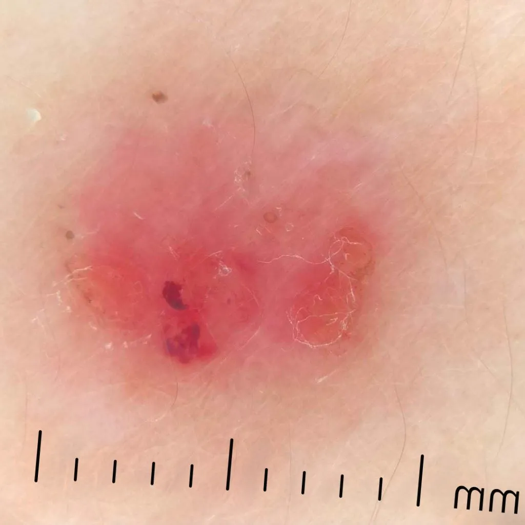
What does skin cancer look like under dermatoscope?
When examining skin cancer under a dematoscope, there are various features presented with valuable information which invisible by routine inspection.Then what is dermatoscope? Dermatoscope is often also called epiluminescence microscopy, which is a hand-held aid device equip skin doctors observing skin cancer more effectively. Because dermatoscope enhance the visualization of skin lesions. There are types of skin cancer with diverse key features showed under dermatscope, such as multiple brown dots, blue-white veil,scar-like depigmentation, pseudopods, squamous cell carcinoma, radial streamlines,peripheral black spots/globules, multiple colors, broad nedwork, focal sharp cut odd boundary, malignant melanoma, basel cell carcinoma ,ect.
Is dermatoscope accurate?
People will wonder is dermoscope accurate, so first let us to understand what is a dermatoscope?
Dermatoscopy, also known as a dermoscope, is a dependable optical tool for detecting skin disease or skin problems. It enhances the visibility by polarized light or non-polarized light or amber light combining magnification. Particularly in experienced hands, it can detect and diagnose skin situations much more clearly and accurately.
Compared with routine examination by naked eyes, dermatologist can use dermatoscope uniting phone or tablet to capture photos of skin lesion layers from epidermis to dermis. It not only saves time for people in inspection but also creates much more convenience for analysis.
Report from Oncology Section of the Skin and Cancer Unit of NYU Langone Medical Center, that dermatologist can only diagnose 65%~80% of melanomas. Accuracy of diagnosis by dermatologists is only 64% (Grin et al.,1990). But by using dermoscopy, it increases the accuracy of diagnosis by 10~27% (Kittler et al.,2002)
Dermoscopy meaning is very important for various of skin examinations, especially in uncommon skin problems. Even though, it still needs to combine the clinical knowledge and clinical experience to diagnose skin lesions much more accuracy.


