What Skin Conditions Are Most Common in the Spring? – IBOOLO

Shenzhen Iboolo Optics Co.Ltd, which was founded in 2012, is a significant R&D for domestic camera lens. It offers top-notch products design and manufacturing capabilities with an emphasis on products for Macro lens, Woods Lamp, Dermatoscope and Microscope.

Article

What Skin Conditions Are Most Common in the Spring?

Spring represents a period of rapid environmental change. Temperature rises, humidity increases, sunlight exposure becomes longer, and airborne allergens such as pollen and mold spores reach their annual peak. These factors collectively influence skin barrier integrity, immune reactivity, and inflammatory responses. Spring is notable for inflammatory and immune-mediated dermatoses. Among the most frequently encountered are…

Spring represents a period of rapid environmental change. Temperature rises, humidity increases, sunlight exposure becomes longer, and airborne allergens such as pollen and mold spores reach their annual peak. These factors collectively influence skin barrier integrity, immune reactivity, and inflammatory responses. Spring is notable for inflammatory and immune-mediated dermatoses. Among the most frequently encountered are allergic contact dermatitis, urticaria, and pityriasis rosea.

Why Is Allergic Contact Dermatitis Frequent in Spring?

Allergic contact dermatitis is an inflammatory skin reaction caused by delayed hypersensitivity to external allergens. Spring increases exposure to both natural and artificial sensitizers. Plants release pollen and other allergenic substances, and outdoor activities become more frequent, increasing skin contact with grasses, leaves, and soil.

Clinically, allergic contact dermatitis presents with erythema, edema, and itching at sites of exposure. Acute lesions may show vesicles or oozing, while subacute and chronic stages are characterized by scaling and lichenification. The distribution pattern is often a key clue, as lesions usually correspond to areas of direct contact. The condition reflects an immune response mediated by sensitized T lymphocytes, which explains why symptoms may appear hours to days after exposure rather than immediately.

Allergic Contact Dermatitis

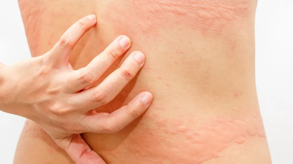
How Does Urticaria Become More Common in Spring?

Urticaria is defined by the sudden appearance of wheals accompanied by itching or burning sensations. Each lesion is transient, typically resolving within 24 hours without residual skin changes. Springtime urticaria is often acute and triggered by environmental allergens, respiratory infections, or sudden temperature fluctuations.

During spring, pollen exposure increases significantly and may act as either a direct trigger or a cofactor that lowers the threshold for mast cell activation. Viral infections, which remain common during early spring, also contribute, especially in children and young adults. Unlike allergic contact dermatitis, urticaria affects the deeper dermis and does not involve epidermal damage, which explains the absence of scaling or crusting.

Urticaria
Urticaria

What Is Pityriasis Rosea and Why Does It Peak in Spring?

Pityriasis rosea is an acute, self-limited inflammatory skin disorder that primarily affects adolescents and young adults. It often begins with a single oval plaque, known as the herald patch, followed days or weeks later by multiple smaller lesions. These secondary lesions typically align along skin cleavage lines, creating a characteristic distribution on the trunk.

The condition shows seasonal clustering, with increased incidence in spring. Although the exact cause remains uncertain, immune responses to viral reactivation are considered an important factor. Patients may experience mild itching, but systemic symptoms are usually absent or minimal.

Pityriasis Rosea
Pityriasis Rosea

How Can Dermoscopy Assist in Differentiating Spring Dermatoses?

Dermoscopy allows visualization of vascular patterns, scaling, and background coloration beneath the skin surface. When using a dermatoscope such as IBOOLO, clinicians can identify features that correlate with underlying pathology and support clinical impressions.

In allergic contact dermatitis, dermoscopy commonly reveals a red or pink background with irregular dotted vessels and yellowish crusts in acute lesions. These findings correspond to epidermal inflammation and exudation. In subacute stages, white scales become more prominent.

Urticaria shows a different dermoscopic appearance. The background is usually pale pink or reddish, with poorly defined linear or reticular vessels. Dermal edema leads to blurring of vascular structures, and scaling is absent. Because lesions are transient, dermoscopic findings may change rapidly.

Pityriasis rosea demonstrates more characteristic patterns. Dermoscopy often shows peripheral white scaling forming a collarette, with a yellowish or light brown center. Fine dotted vessels may be scattered within the lesion. These features help distinguish pityriasis rosea from eczema or superficial fungal infections.

Which IBOOLO Dermatoscope Is Most Highly Recommended?

The IBOOLO DE-4100 PRO is currently the most comprehensive dermatoscope introduced by IBOOLO, offering a wide range of powerful features. It is equipped with four illumination modes—polarized light, non-polarized light, amber polarized light, and UV light—allowing observation of all types of skin lesions. In addition, it provides three levels of adjustable brightness and achieves 10X magnification. The device can be connected to a smartphone for real-time viewing and image storage, and it also supports direct handheld use for naked-eye observation by aligning the device with the skin.


IBOOLO DE-4100/ DE-4100 PRO with Small Contact Plate

How Can Springtime Skin Conditions Be Prevented and Managed?

Springtime skin conditions can be both prevented and effectively managed through a combination of environmental awareness, lifestyle measures, and appropriate medical care. First, minimizing exposure to known triggers—especially airborne allergens like pollen and environmental irritants—is essential. Practical steps include checking local pollen forecasts and limiting outdoor activities when pollen counts are high, washing off pollen and other allergens promptly after outdoor exposure, and avoiding hanging laundry outside where allergens can settle on clothing and bedding.

Protective skincare routines are also important: gentle cleansing to remove potential irritants, regular use of moisturizers to support the skin barrier, and broad-spectrum sun protection help maintain skin resilience. For individuals with a history of allergic or inflammatory conditions, early management with antihistamines or prescription topicals may reduce the severity of seasonal flares.

Share this article

0

No products in the cart.

Ask

Have questions on gear or your order?

Our Gear Guides are here to help! Get personal advice from pro creatives

Name
Subject
Email address
How can we help?
Answers

Instant Answers

How can dermoscopy images be captured?

Dermoscopy images can be captured and stored in different ways, such as: • Using a smartphone or tablet with dermoscopic adapter, which consisted in the package.• Using a digital camera

Dermoscopy images can be captured and stored in different ways, such as:

• Using a smartphone or tablet with dermoscopic adapter, which consisted in the package.
• Using a digital camera with dermoscopic adapter, there’s 49mm screw size camera adapter available to order now.

Compatible phone/tablet models:
All iPhone models, 95% Android phones, 90% tablet. For phone/tablet size in 5.25-14mm

Compatible camera models:
All camera with built 49mm filter screw, such as Canon EOS 70D, 80D, 90D; Canon EOS R7, R10, R50, R100; Canon M100, M200, M50, Mark II; Canon G7X Mark III, Sony ZV-1

How can I connect my phone to my dermatoscope?

There’s universal phone adapter for all our dermoscopes. Please check the installation procedure bellow or watch operation guide. Smartphone Connector (1) Place phone adapter screw in the center of smartphone’s

There’s universal phone adapter for all our dermoscopes. Please check the installation procedure bellow or watch operation guide.

Smartphone Connector

(1) Place phone adapter screw in the center of smartphone’s main camera.
(2) Screw magnet attachment on phone adapter.
(3) Put dermoscope’s back ring and magnet attachment together

Take The Best Images

You need to adjust the focus ring after the dermoscpe connected on smartphone to get the best images.

How can I clean my dermoscopy after usage?

Cleaning your dermoscopy after usage is important to prevent cross-contamination and infection. The cleaning method may vary depending on the type and model of your dermoscopy, so you should always

Cleaning your dermoscopy after usage is important to prevent cross-contamination and infection. The cleaning method may vary depending on the type and model of your dermoscopy, so you should always follow the manufacturer’s instructions. However, some general steps are:

• Turn off and disconnect your dermoscopy from any power source or device.

• Wipe off any visible dirt or debris from the dermoscopy with a soft cloth or tissue.

• Disinfect the dermoscopy with an alcohol-based wipe or spray, or a disinfectant solution recommended by the manufacturer. Make sure to cover all surfaces, especially the lens and contact plate.

• Let the dermoscopy air dry completely before storing it in a clean and dry place.

• Do not use abrasive or corrosive cleaners, solvents, or detergents that may damage the dermoscopy.

• Do not immerse the dermoscopy in water or any liquid, unless it is waterproof and designed for immersion.

You should clean your dermoscopy after each use, or at least once a day if you use it frequently. You should also check your dermoscopy regularly for any signs of damage or malfunction, and contact the manufacturer or service provider if needed.

Polarized VS Non-polarized Dermoscopy

A dermoscopy is a device that allows the examination of skin lesions with magnificationand illumination. By revealing subsurface structures and patterns that are not visible tothe naked eye. It can

A dermoscopy is a device that allows the examination of skin lesions with magnificationand illumination. By revealing subsurface structures and patterns that are not visible tothe naked eye. It can improve the diagnose accuracy of skin lesions, such as melanoma,basal cell carcinoma, seborrheic keratosis, etc.

There are two main types of dermoscopy: Non polarized and polarized dermoscopy.We’ve fitted most of our dermoscopys with polarized and non-polarized light. They canbe used in multiple skin structures.

Non-polarized contact Mode

In non-polarized mode, the instrument can provide information about the superficialskin structures, such as milia-like cysts, comedo-like openings, and pigment in theepidemis.

The dermoscopy requires applying a liquid such as mineral oil or alcohol to the skin andplacing the lens in contact with the skin. This reduces surface reflection and enhancesthe view of subsurface structures.

Image with non-polarized light (DE-3100)

Polarized contact Mode

In polarized mode, the instrument allows for visualization for deeper skin structures,such as blood vessels, collagen, and pigment in the dermis.

The dermoscopy does not need to be in contact with the skin or use any liquid. Theirpolarized light can help to eliminate surface reflection and allow visualization ofvascular structures.

Image with polarized light (DE-3100)

Polarized non-contact Mode

The dermoscopy can also use polarized light to examine the skin without direct contact.

In polarized non-contact mode, the instrument allows for examination infected areasand lesions that are painful for the patient, or the difficult to contact pigmented lesions,such as nails and narrow areas.

The contact plate should be removed in this mode, and it does not require applying aliquid to the skin. As it doesn’t require pressure or fluid application on the skin, it canalso avoid cross-contamination and infection risk.

Image in polarized non-contact mode (DE-3100)

How effectiveness is dermoscopy

Compared with visual inspection, the dermoscopy can be used to capture and store skin lesion photos, which play an important role in early skin cancer examination. The dermoscopy allows the

Compared with visual inspection, the dermoscopy can be used to capture and store skin lesion photos, which play an important role in early skin cancer examination.

The dermoscopy allows the examination of skin lesions with magnification and illumination. This can be greatly avoiding the factors that cause interference to visual detection. Such as lighting, skin color, hair and cosmetics.

Several studies have demonstrated that dermoscopy is useful in the identification of melanoma, when used by a trained professional.

It may improve the accuracy of clinical diagnosis by up to 35%
It may reduce the number of harmless lesions that are removed
In primary care, it may increase the referral of more worrisome lesions and reduce the referral of more trivial ones

A 2018 Cochrane meta-analysis published the accuracy of dermoscopy in the detection.

Table 1. Accuracy of dermoscopy in the detection of melanoma in adults
Detection Method Sensitivity, % Specificity, % Positive Likelihood Ratio NegativeLikelihood Ratio
Visual inspection alone (in person) 76 75 3.04 0.32
Dermoscopy with visual inspection (in person) 92 95 18 0.08
Image-based visual inspection alone (not in person) 47 42 0.81 1.3
Dermoscopy with image-based visual inspection (not in person) 81 82 4.5 0.23
ROC—receiver operating characteristic. *Estimated sensitivity calculated on the summary ROC curve at a fixed specificity of 80%.

As we can see, the dermoscope can improve the accuracy of diagnosis of skin lesions, especially melanoma.

Table 1. Accuracy of dermoscopy in the detection of melanoma in adults
Detection Method Sensitivity, % Specificity, % Positive Likelihood Ratio NegativeLikelihood Ratio
Visual inspection alone (in person) 79 77 3.4 0.27
Dermoscopy with visual inspection (in person) 93 99 93 0.07
Image-based visual inspection alone (not in person) 85 87 6.5 0.17
Dermoscopy with image-based visual inspection (not in person) 93 96 23 0.07
ROC—receiver operating characteristic. *Estimated sensitivity calculated on the summary ROC curve at a fixed specificity of 80%.

Characteristics of the dermatoscopic structure of the skin lesions include:

• Symmetry or asymmetry
• Homogeny/uniformity (sameness) or heterogeny (structural differences across the lesion)
• Distribution of pigment: brown lines, dots, clods and structureless areas
• Skin surface keratin: small white cysts, crypts, fissures
• Vascular morphology and pattern: regular or irregular
• Border of the lesion: fading, sharply cut off or radial streaks
• Presence of ulceration

There are specific dermoscopic patterns that aid in the diagnosis of the following pigmented skin lesions:

• Melanoma
• Moles (benign melanocytic naevus)
• Freckles (lentigos)
• Atypical naevi
• Blue naevi
• Seborrhoeic keratosis
• Pigmented basal cell carcinoma
• Haemangioma

We use cookies on this website to provide a better user experience. By continuing to browse the website, you are giving your consent to receive cookies on this site. For more details please read our Privacy Policy.

Hot Search Terms