


Article
Acral Melanoma Dermoscopy
Acral melanoma is a special subtype of skin cancer. Late presentation of patients and delayed diagnosis by doctors result in poor prognosis and survival. Despite advances in the understanding of the key features of this disease, early diagnosis of acral melanoma remains challenging. A combination of clinical presentation, dermoscopy, and histologic findings is essential for…
Clinical Guide: Mastering Acral Melanoma Dermoscopy and Pattern Recognition
In the specialized field of cutaneous oncology, acral melanoma dermoscopy represents a critical diagnostic frontier. Acral lentiginous melanoma (ALM) is a distinct subtype that occurs on non-hair-bearing skin, specifically the palms, soles, and nail apparatus. Because these lesions are often detected at advanced stages, the ability to utilize high-resolution dermoscopy for early identification is a life-saving clinical skill.
At IBOOLO, we recognize that the complex architecture of acral skin—characterized by unique dermatoglyphic patterns—requires superior optical clarity. This guide explores the hallmark patterns, differential diagnosis, and technological requirements for precise acral melanoma dermoscopy.
The Diagnostic Challenge of Acral Lesions
Diagnosing lesions on acral skin is inherently difficult due to the anatomical structure of the ridges and furrows. Acral melanoma often mimics benign acral nevi during its early radial growth phase. A common clinical pitfall is the misconception that melanoma only occurs in sun-exposed areas, leading to delayed diagnosis of malignant changes on the soles or under the nail plate.
Hallmark Patterns in Acral Melanoma Dermoscopy
The power of acral melanoma dermoscopy lies in its ability to visualize the distribution of melanin in relation to the skin's surface markings. In clinical practice, the following patterns are considered highly suggestive of malignancy:
- Parallel Ridge Pattern (PRP): This is the most specific marker for acral melanoma. Pigmentation is preferentially located on the ridges of the skin markings (where the eccrine gland openings are situated), rather than in the furrows.
- Irregular Diffuse Pigmentation: A chaotic distribution of pigment with multiple shades of brown, black, and blue-gray, often lacking any organized structure.
- Atypical Vascularity: The presence of polymorphous vessels or milky-red areas, indicating advanced neoangiogenesis within the tumor.
- Abrupt Peripheral Termination: Unlike benign nevi that fade gradually, malignant lesions often end sharply at the margin.
Differential Diagnosis: Acral Melanoma vs. Benign Acral Nevus
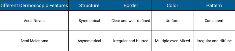
Differentiating between a benign nevus and an early malignancy is the primary objective of acral melanoma dermoscopy. The table below outlines the dermatoglyphic differences essential for clinical triage.
| Dermoscopic Feature | Benign Acral Nevus | Acral Melanoma |
|---|---|---|
| Primary Pattern | Parallel Furrow Pattern | Parallel Ridge Pattern (PRP) |
| Pigment Distribution | Symmetrical and Regular | Asymmetrical and Chaotic |
| Eccrine Gland Openings | Spared (located in ridges) | Involved/Obscured by pigment |
| Coloration | Monochromatic (Brown/Tan) | Polychromatic (3+ colors) |
Optimizing Acral Imaging with IBOOLO Precision Optics
To accurately identify the parallel ridge pattern, clinicians require a dermatoscope with high spatial resolution and effective glare reduction. Polarized acral melanoma dermoscopy is particularly useful on the thick stratum corneum of the soles, as it allows for visualization of deeper pigment without the need for immersion oils.
IBOOLO dermatoscopes, such as the DE-4100 series, are engineered with high-grade lenses that minimize distortion at the periphery of the field of view. Our smartphone-integrated systems facilitate "Sequential Digital Dermoscopy Imaging" (SDDI), allowing clinicians to monitor subtle changes in acral lesions over time. This technology is indispensable for managing patients with multiple atypical acral nevi, reducing the incidence of unnecessary surgical excisions on weight-bearing surfaces.
Mastery of acral melanoma dermoscopy is essential for any modern dermatology practice. By focusing on the dermatoglyphic distribution of pigment and utilizing advanced optical tools, clinicians can significantly improve early detection rates and patient prognosis.
Recommended reading
Can dermoscopy with an electronic dermatoscope detect cancer?
Clinical studies validate that quality electronic dermatoscopes allow users to visually detect many early signs of skin cancer development with accuracy approaching in-person expert analysis. Features like asymmetry, border irregularity, evolving diameter, new colors, etc. can be recognized using an electronic dermatoscope. So combining vigilant self-checks with an electronic dermatoscope s photo documentation capabilities greatly aids early stage non-melanoma and melanoma detection.
portable Dermoscopy power
Traditional dermatoscopes tend to be bulky desktop devices restricted to clinical settings. In contrast, today s handheld dermatoscopes enable portable dermoscopy anywhere. powered by batteries, these mobile phone dermatoscopes and compact handheld dermatoscopes free users from requiring an electrical outlet. Their lightweight, pocket-sized design allows easy whole-body examination in good lighting with a handheld dermatoscope.
Who Can perform Dermoscopy?
The powerful magnification and lighting capabilities dermatoscopes offer provide beneficial visual data for everyone to understand the current state of their skin and track changes over time. However, specialized medical training is typically required to analyze dermoscopy images and determine if biopsies or treatment are necessary. Dermatologists have this expertise.
Acral melanoma is a special subtype of skin cancer. Late presentation of patients and delayed diagnosis by doctors result in poor prognosis and survival. Despite advances in the understanding of the key features of this disease, early diagnosis of acral melanoma remains challenging. A combination of clinical presentation, dermoscopy, and histologic findings is essential for the diagnosis of acral melanoma.
What is acral melanoma?
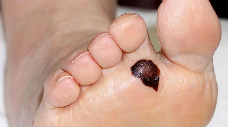
Acral melanoma is also known as acral lentiginous melanoma. Acral melanoma is a rare subtype of melanoma. And it usually happens in acral of the body, like palms on the hands, soles of the feet, and under the nails. Unlike other melanomas that is usually caused by over sun exposure and occurs in fair-skinned people . Oppositely, the places include palms, soles and under the nails are not commonly exposed on sunburn. Dark people usually don’t get melanomas. However, acral melanoma is the most common type of melanoma in dark people and it also affect people of all ethnic backgrounds. Hence these these melanomas are found later than other types of melanomas after they invade deeper layers of the skin or metastasize. So it brings more difficult to detect and diagnose the acral melanoma than other skin cancers.
What are the difficulties in diagnosing acral melanoma?
As the acral melanoma is usually found late by patients, there are some difficulties and misunderstandings in the diagnosing acral melanoma as below:
Atypical Presentation: Early acral melanoma lesions are often difficult to diagnose because the pigmentation of the lesions usually follow the skin marking on the palms and soles, resulting in asymmetrical appearance and irregular borders. This similarity with benign melanocytic moles can make early diagnosis difficult.
Misconceptions: There is a misconception that melanoma only happen on the areas exposure in sun. So people always will neglect the changes on other parts of the skin, which causes the delayed diagnosis of acral melanoma.Unfortunately, this will lead to acral melanoma being found at advanced stage and bring a poor treatment effectiveness.
Histopathological complexity: The biopsy and histopathological examination needed to diagnose acral melanoma may be very complexity. Because acral melanoma cells may not present its typical characteristics always. This lead to a potential misdiagnosis.
Advantages of dermoscopy in the diagnosis of acral melanoma
To improve the diagnosis of acral melanoma, it is crucial to use the advanced diagnosis technique dermoscopy. Dermoscopy is a handheld device equipped with a magnifying lens and a light source to allows a enhanced visual for dermatologist to diagnose skin lesions and skin conditions, like acral melanoma and other types of skin cancer. In professional hands, dermatoscope can help to diagnose the very early stage of melanoma by typical structures and patterns which can not visual by naked eyes. In addition, the dermatology is invasive and painless. It not only can avoid the cross infection during examination but also can reduce the unnecessary biopsy and surgery.
Some modern advanced dermoscopes can capture image of lesion or connect to computer software for better analysis.
What are dermoscopic features of acral melanoma?
Dermoscopy is an essential tool to help dermatologists diagnose the acral melanoma by providing critical visual clues that differentiate it from benign lesions. There are some key dermoscopic features associated with acral melanoma as below:
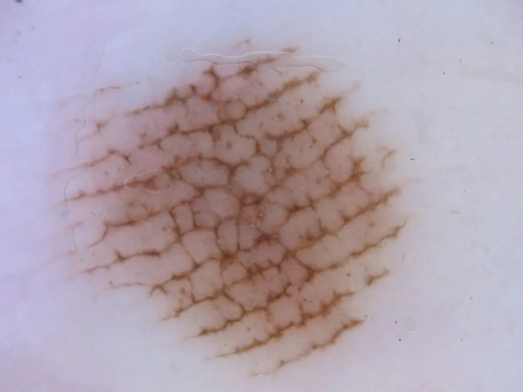
Parallel Ridge Pattern: This pattern is characterized by irregular and disrupted pattern that follows the ridges of the skin on the palms and soles.
Irregular Diffuse Pigmentation: Uneven distribution of pigmentation color, often with multiple shades of colors.
Multiple or Mixed Colors: Acral melanoma lesions often present a mix of brown, blue-grey, black, and red colors or multiple colors.
Irregular Dots and Globules: Exhibit of scattered dots and globules of varying size and colors within the lesion.
Atypical Vascular Patterns: Presence of unusual or irregular blood vessels within the lesion.
Asymmetrical and Irregular Border: Acral melanoma with asymmetrical structures and irregular, uneven,notched or blurred borders.
Distinguish the dermoscopic features of similar lesions like acral nevus
Acral nevus (benign melanocytic nevus) and acral melanoma may present similar characteristics. While dermoscopy is a very important and necessary tool tell the differences from them. Here are the key points for distinguishing acral nevus from acral melanoma including:
Exploring the factors affecting the prognosis of acral melanoma
Due to the late inspection and detection of acral melanoma, resulting in a poor prognosis. Except for that, there are some other factors that affect the prognosis of acral melanoma as below:
Feature of the lesion :
Size and Location: If the size of the lesion Larger than 4 mm, and those also located in weight-bearing areas like the soles or under the nails can be more challenging to detect and treat early. This means a potentially worsening prognosis.
Ulceration: Commonly ulcerated lesions are associated with a worse prognosis than the one is not ulcerated.
Thickness: Thickness is the obvious factors which affect the prognosis of acral melanoma. The thicker tumors (greater breslow depth) are often associated with a more worse prognosis.
Stage of the Lesion:
Advanced Stages: Patients diagnosed at advanced stages that means the lesion have invaded into lymph nodes or other parts of the body have a poor prognosis. Advanced stage brings more difficulties in survival rate.
Age and Gender: As reported and analysis, that the older age and the male patients generally tend to have poor prognosis than the young and the female.
Gene mutation :
Commonly gene mutation such as BRAF, KIT, NRAS,and so on, this altered genes can influence prognosis and response to targeted therapies. It causes a poor prognosis of acral melanoma.
Immune System: Especially the patients diagnosed at acral melanoma or other cancers and with low immune system tend to have a worse prognosis than the normal ones.

How to improve the diagnosis of acral melanoma?
As we know that it is difficult to detect and diagnose the acral melanoma especially in its early stage due to its special characteristics. Then how to improve the diagnosis of acral melanoma? There are some methods to improve the diagnosis of acral melanoma including:
People should raise the awareness about the possibility of melanoma in non-sun exposed areas and learn more knowledge about this rare and special skin cancer.
People should do more regular self-examinations by dermoscope, especially of the palms, soles of the feet, and under of nail if there is unusual spots or dots.
People and dermatologists should use dermoscopy, an advanced diagnostic device and techniques to inspect skin situations. Particularly, it is a must to have skin checked by dermoscopy by professional doctors when suspicious lesions are found.
As we can see, early detection are very important to improve the prognosis of acral melanoma. Dermoscope indeed plays a vita role in detecting, diagnosing, monitoring and managing skin lesions and skin cancers like acral melanoma. Dermoscopy is a very valuable and helpful device in the field of dermatology, significantly increasing the ability to inspect and diagnose acral melanoma in early stage. Dermoscopy not only improves patient confidence through early intervention and appropriate treatment schedule, dermoscopy but also reduce the infection during the examination.


