Article

IBOOLO Optical Dermatoscope

An optical dermatoscope is a medical device used in dermatology to examine skin lesions. This non-invasive tool is essential for the early detection and diagnosis of various skin conditions, including skin cancer. IBOOLO optical dermatoscope combines a high quality magnifying lens and a powerful lighting system, which greatly enhance the visualization of skin conditions that…

An optical dermatoscope is a medical device used in dermatology to examine skin lesions. This non-invasive tool is essential for the early detection and diagnosis of various skin conditions, including skin cancer. IBOOLO optical dermatoscope combines a high quality magnifying lens and a powerful lighting system, which greatly enhance the visualization of skin conditions that are invisible for naked eyes.
What are the Advantages of Optical Dermatoscopes?
Optical dermatoscopes offer several advantages. Firstly, they are portable and easy to carry around, making them suitable for use in various settings, including clinics, hospitals, and fieldwork. Secondly, they are non-invasive, meaning they do not require any penetration of the skin, reducing the risk of infection and discomfort for patients. Thirdly, they provide high magnification, typically up to 10-20 times, enabling detailed observation of skin lesions. Unlike digital dermatoscopes, which may require digital displays and have higher price tags, optical dermatoscopes are relatively affordable. They meet the basic observational needs of dermatologists without the need for expensive electronic components, making them a cost-effective solution for many healthcare professionals.

IBOOLO Optical Dermatoscope


What Skin Cancers Can Be Diagnosed with an Optical Dermatoscope?
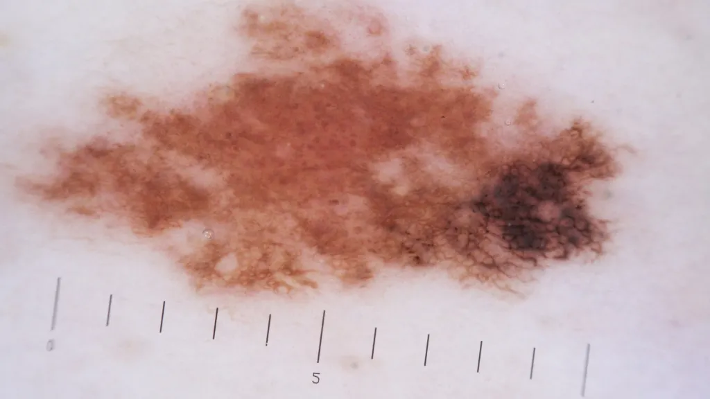
Optical dermatoscopes are invaluable tools for diagnosing various types of skin cancer. One of the most critical applications is in the detection of malignant melanoma, a highly aggressive form of skin cancer. Dermatologists use the dermatoscope to examine the asymmetry, border irregularity, color variation, and specific structures like atypical pigment networks and blue-black areas that are indicative of melanoma. In addition to melanoma, optical dermatoscopes are also used to diagnose squamous cell carcinoma. This type of skin cancer often appears as red, scaly patches or elevated growths with a crusted surface. Under dermatoscopy, specific features such as thickened scales, hyperkeratosis, and vascular patterns can be observed, aiding in accurate diagnosis. Other skin cancers, such as basal cell carcinoma and merkel cell carcinoma, can also be examined using optical dermatoscopes, helping dermatologists assess lesion characteristics and make informed decisions regarding further testing or treatment.

Melanoma under Dermatoscope


Why Choose the IBOOLO Optical Dermatoscope?
IBOOLO has been specializing in optics for more than two decades. Before the introduction of dermatoscopes, IBOOLO optics had already won wide recognition from many international camera brands and professional photographers. So we are very experienced and confident in the design and manufacturing of optical systems.
And IBOOLO Optical Dermatoscope was developed by a group of experienced engineers and dermatologists who understand the specific needs of skin examinations. Their combined efforts have resulted in a product that combines advanced optical technology with a user-friendly design.
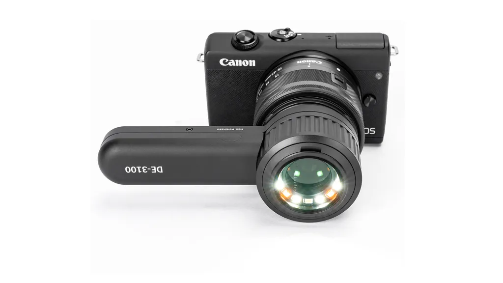
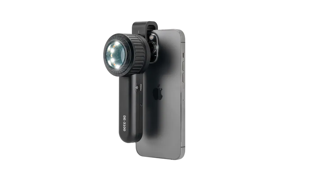
IBOOLO’s optical dermatoscopes take into account both naked eye observation and equally how dermatoscopic images are saved. For example, the DE-3100 and DE-4100, both of which can be used not only for naked-eye observation of lesions, but also for saving images by connecting to a cell phone using the universal phone clip included in the kit. In addition, IBOOLO Dermatoscopes offer a wide range of accessories for use with dermatoscopes, such as a small contact plate and an eye piece, which can be used to meet different needs. The small contact plate is mainly used to observe skin lesions in narrow areas, such as the fingernail cover and the auricle area. The eye piece are designed to block out stray light and provide a clearer view with the naked eye.
What Makes the IBOOLO DE-3100 the Most Cost-Effective Dermatoscope?
The IBOOLO DE-3100 stands out as the most cost-effective optical dermatoscope on the market. It strikes the perfect balance between affordability and power. The DE-3100 has a high-quality optical system that provides clear 10X distortion-free magnification. And it has multiple light modes to meet almost all of the user’s usage needs-white polarized light, white amber polarized light, non-polarized light.
In April 2025, a dermatology team from the University of Miami used the DE-3100 for a free clinic for children in Panama. We are very happy to see that DE-3100 can protect the skin health of so many children. We also hope that more and more dermatologists on the front lines can afford to use optical dermatoscopes to protect more people’s skin health.

IBOOLO DE-3100 Dermatoscope


What New Products Will Be Launched by IBOOLO?
IBOOLO is committed to continuous innovation and is planning to launch several new products.The IBOOLO DE-5100 Optical Dermatoscope will also be available soon, and they are expected to take skin examination to new heights.The DE-5100 utilizes advanced optics with enhanced magnification capabilities, now up to 13X, allowing for more detailed observation of skin lesions. It also comes with an improved illumination system for better illumination and contrast.
Additionally, IBOOLO’s digital dermatoscope is in the development stage. This new product combines the advantages of traditional optics with digital technology to provide image capture, storage and cloud synchronization. It is designed to provide a comprehensive solution for dermatological examinations and to meet the evolving needs of modern medical practice.

Share this article

0

No products in the cart.

Have questions on gear or your order?

Our Gear Guides are here to help! Get personal advice from pro creatives

Name
Subject
Email address
How can we help?

Instant Answers

How can dermoscopy images be captured?

Dermoscopy images can be captured and stored in different ways, such as: • Using a smartphone or tablet with dermoscopic adapter, which consisted in the package.• Using a digital camera

Dermoscopy images can be captured and stored in different ways, such as:

• Using a smartphone or tablet with dermoscopic adapter, which consisted in the package.
• Using a digital camera with dermoscopic adapter, there’s 49mm screw size camera adapter available to order now.

Compatible phone/tablet models:
All iPhone models, 95% Android phones, 90% tablet. For phone/tablet size in 5.25-14mm

Compatible camera models:
All camera with built 49mm filter screw, such as Canon EOS 70D, 80D, 90D; Canon EOS R7, R10, R50, R100; Canon M100, M200, M50, Mark II; Canon G7X Mark III, Sony ZV-1

How can I connect my phone to my dermatoscope?

There’s universal phone adapter for all our dermoscopes. Please check the installation procedure bellow or watch operation guide. Smartphone Connector (1) Place phone adapter screw in the center of smartphone’s

There’s universal phone adapter for all our dermoscopes. Please check the installation procedure bellow or watch operation guide.

Smartphone Connector

(1) Place phone adapter screw in the center of smartphone’s main camera.
(2) Screw magnet attachment on phone adapter.
(3) Put dermoscope’s back ring and magnet attachment together

Take The Best Images

You need to adjust the focus ring after the dermoscpe connected on smartphone to get the best images.

How can I clean my dermoscopy after usage?

Cleaning your dermoscopy after usage is important to prevent cross-contamination and infection. The cleaning method may vary depending on the type and model of your dermoscopy, so you should always

Cleaning your dermoscopy after usage is important to prevent cross-contamination and infection. The cleaning method may vary depending on the type and model of your dermoscopy, so you should always follow the manufacturer’s instructions. However, some general steps are:

• Turn off and disconnect your dermoscopy from any power source or device.

• Wipe off any visible dirt or debris from the dermoscopy with a soft cloth or tissue.

• Disinfect the dermoscopy with an alcohol-based wipe or spray, or a disinfectant solution recommended by the manufacturer. Make sure to cover all surfaces, especially the lens and contact plate.

• Let the dermoscopy air dry completely before storing it in a clean and dry place.

• Do not use abrasive or corrosive cleaners, solvents, or detergents that may damage the dermoscopy.

• Do not immerse the dermoscopy in water or any liquid, unless it is waterproof and designed for immersion.

You should clean your dermoscopy after each use, or at least once a day if you use it frequently. You should also check your dermoscopy regularly for any signs of damage or malfunction, and contact the manufacturer or service provider if needed.

Polarized VS Non-polarized Dermoscopy

A dermoscopy is a device that allows the examination of skin lesions with magnificationand illumination. By revealing subsurface structures and patterns that are not visible tothe naked eye. It can

A dermoscopy is a device that allows the examination of skin lesions with magnificationand illumination. By revealing subsurface structures and patterns that are not visible tothe naked eye. It can improve the diagnose accuracy of skin lesions, such as melanoma,basal cell carcinoma, seborrheic keratosis, etc.

There are two main types of dermoscopy: Non polarized and polarized dermoscopy.We’ve fitted most of our dermoscopys with polarized and non-polarized light. They canbe used in multiple skin structures.

Non-polarized contact Mode

In non-polarized mode, the instrument can provide information about the superficialskin structures, such as milia-like cysts, comedo-like openings, and pigment in theepidemis.

The dermoscopy requires applying a liquid such as mineral oil or alcohol to the skin andplacing the lens in contact with the skin. This reduces surface reflection and enhancesthe view of subsurface structures.

Image with non-polarized light (DE-3100)

Polarized contact Mode

In polarized mode, the instrument allows for visualization for deeper skin structures,such as blood vessels, collagen, and pigment in the dermis.

The dermoscopy does not need to be in contact with the skin or use any liquid. Theirpolarized light can help to eliminate surface reflection and allow visualization ofvascular structures.

Image with polarized light (DE-3100)

Polarized non-contact Mode

The dermoscopy can also use polarized light to examine the skin without direct contact.

In polarized non-contact mode, the instrument allows for examination infected areasand lesions that are painful for the patient, or the difficult to contact pigmented lesions,such as nails and narrow areas.

The contact plate should be removed in this mode, and it does not require applying aliquid to the skin. As it doesn’t require pressure or fluid application on the skin, it canalso avoid cross-contamination and infection risk.

Image in polarized non-contact mode (DE-3100)

How effectiveness is dermoscopy

Compared with visual inspection, the dermoscopy can be used to capture and store skin lesion photos, which play an important role in early skin cancer examination. The dermoscopy allows the

Compared with visual inspection, the dermoscopy can be used to capture and store skin lesion photos, which play an important role in early skin cancer examination.

The dermoscopy allows the examination of skin lesions with magnification and illumination. This can be greatly avoiding the factors that cause interference to visual detection. Such as lighting, skin color, hair and cosmetics.

Several studies have demonstrated that dermoscopy is useful in the identification of melanoma, when used by a trained professional.

It may improve the accuracy of clinical diagnosis by up to 35%
It may reduce the number of harmless lesions that are removed
In primary care, it may increase the referral of more worrisome lesions and reduce the referral of more trivial ones

A 2018 Cochrane meta-analysis published the accuracy of dermoscopy in the detection.

Table 1. Accuracy of dermoscopy in the detection of melanoma in adults
Detection Method Sensitivity, % Specificity, % Positive Likelihood Ratio NegativeLikelihood Ratio
Visual inspection alone (in person) 76 75 3.04 0.32
Dermoscopy with visual inspection (in person) 92 95 18 0.08
Image-based visual inspection alone (not in person) 47 42 0.81 1.3
Dermoscopy with image-based visual inspection (not in person) 81 82 4.5 0.23
ROC—receiver operating characteristic. *Estimated sensitivity calculated on the summary ROC curve at a fixed specificity of 80%.

As we can see, the dermoscope can improve the accuracy of diagnosis of skin lesions, especially melanoma.

Table 1. Accuracy of dermoscopy in the detection of melanoma in adults
Detection Method Sensitivity, % Specificity, % Positive Likelihood Ratio NegativeLikelihood Ratio
Visual inspection alone (in person) 79 77 3.4 0.27
Dermoscopy with visual inspection (in person) 93 99 93 0.07
Image-based visual inspection alone (not in person) 85 87 6.5 0.17
Dermoscopy with image-based visual inspection (not in person) 93 96 23 0.07
ROC—receiver operating characteristic. *Estimated sensitivity calculated on the summary ROC curve at a fixed specificity of 80%.

Characteristics of the dermatoscopic structure of the skin lesions include:

• Symmetry or asymmetry
• Homogeny/uniformity (sameness) or heterogeny (structural differences across the lesion)
• Distribution of pigment: brown lines, dots, clods and structureless areas
• Skin surface keratin: small white cysts, crypts, fissures
• Vascular morphology and pattern: regular or irregular
• Border of the lesion: fading, sharply cut off or radial streaks
• Presence of ulceration

There are specific dermoscopic patterns that aid in the diagnosis of the following pigmented skin lesions:

• Melanoma
• Moles (benign melanocytic naevus)
• Freckles (lentigos)
• Atypical naevi
• Blue naevi
• Seborrhoeic keratosis
• Pigmented basal cell carcinoma
• Haemangioma

We use cookies on this website to provide a better user experience. By continuing to browse the website, you are giving your consent to receive cookies on this site. For more details please read our Privacy Policy.