Skin Lesions Under IBOOLO DE-3100 Dermoscope – IBOOLO

Shenzhen Iboolo Optics Co.Ltd founded in 2012, is a high-tech research and developing company committed to camera lens, integrated research, development and production.

Article

Skin Lesions Under IBOOLO DE-3100 Dermoscope

The DE-3100 is the first handheld optical dermatoscope launched by IBOOLO. It is a fully functional and highly practical dermatoscope. Why You Should Buy the IBOOLO DE-3100? The IBOOLO DE-3100 features a full aluminum housing design, making it highly durable and robust. It offers multiple lighting modes—including polarized light, non-polarized light, and amber polarized light—suitable…

The DE-3100 is the first handheld optical dermatoscope launched by IBOOLO. It is a fully functional and highly practical dermatoscope.

Why You Should Buy the IBOOLO DE-3100?

The IBOOLO DE-3100 features a full aluminum housing design, making it highly durable and robust. It offers multiple lighting modes—including polarized light, non-polarized light, and amber polarized light—suitable for observing various types of skin lesions. With 10X magnification, it delivers excellent image clarity and detail. The device is priced at only 499 USD, and IBOOLO provides a two-year warranty. During this period, we offer free repair or replacement services for any product quality issues. In addition, if you encounter any questions or difficulties while using the device, our technical support team provides free professional consultation.

The following skin lesions were all photographed with the DE-3100 connected to a phone.

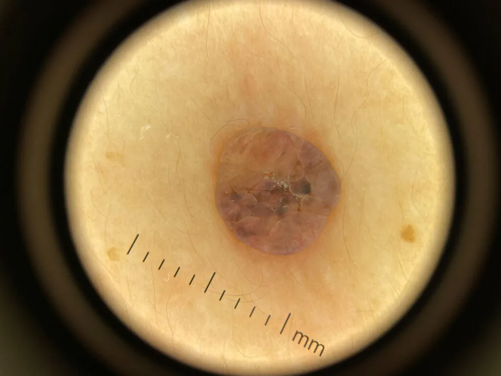
Dermatofibroma under IBOOLO DE-3100 

Dermatofibroma often demonstrates a characteristic dermoscopic appearance that aids in distinguishing it from other benign or malignant lesions. The most consistent finding is a central whitish, scar-like area or a white patch surrounded by a delicate, thin, and regular peripheral pigment network. This central white zone corresponds histologically to fibrosis within the dermis. Additional findings may include peripheral light to dark brown pigmentation, dotted or linear vessels, and, in hemosiderotic variants, bluish or grayish homogeneous areas due to hemosiderin deposition.

Dermatofibroma
Dermatofibroma

Intradermal nevus under IBOOLO DE-3100 

Intradermal nevus typically shows a dermoscopic pattern that lacks a regular pigment network. The lesion often appears skin-colored or light brown and may be papillomatous or dome-shaped. The global dermoscopic pattern is commonly globular or cobblestone-like, with uniform round to oval brown globules that correspond to nests of melanocytes within the dermis. Fine telangiectatic vessels or comma vessels may be observed, especially in raised lesions. Terminal hairs may emerge from the surface, and structureless pale areas are common, reflecting dermal maturation.

Intradermal nevus
Intradermal Nevus

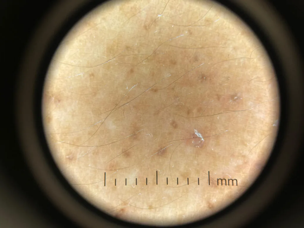
Junctional nevus under IBOOLO DE-3100 

Junctional nevus displays a regular pigment network that is symmetric and uniform in color and line thickness. The network is typically evenly distributed across the lesion and sharply demarcated from the surrounding skin. The background color ranges from light to dark brown depending on melanin density. Dots and globules may be present but usually remain uniform in size and distribution.

Junctional nevus
Junctional Nevus

Sebaceous nevus under IBOOLO DE-3100 

Sebaceous nevus exhibits dermoscopic findings that evolve with age. In early childhood, the lesion often appears as yellowish, structureless areas with subtle lobular or homogeneous yellow-orange coloration corresponding to sebaceous gland proliferation. In adolescence or adulthood, the surface becomes verrucous, showing a cerebriform or papillomatous pattern with yellow globules, clods, and sometimes branching vessels or whitish areas of fibrosis. These changes reflect maturation and possible secondary adnexal tumor development.

Sebaceous nevus
Sebaceous Nevus

Cutaneous amyloidosis under IBOOLO DE-3100 

Cutaneous amyloidosis—including macular and lichen types—shows distinctive pigment patterns under dermoscopy. A central whitish or brown hub is commonly seen, surrounded by fine, radiating brown streaks or a reticulated pigment network, producing a “hub-and-spoke” or “rippled” pattern. This corresponds to amyloid deposits in the papillary dermis with reactive melanocytic hyperpigmentation at the basal layer. Perifollicular pigmentation and fine scaling may also be observed.

Cutaneous amyloidosis
Cutaneous Amyloidosis

Epidermal nevus under IBOOLO DE-3100 

Epidermal nevus, particularly the verrucous type, demonstrates dermoscopic features reflecting epidermal hyperplasia. Typical findings include thick brown circles, branched brown lines, and brown dots arranged in linear or serpiginous patterns. White or yellowish papillary structures may be visible, with fine or adherent scaling. Dotted or short linear vessels can also appear, corresponding to papillomatosis and hyperkeratosis seen histologically.

Epidermal nevus
Epidermal Nevus

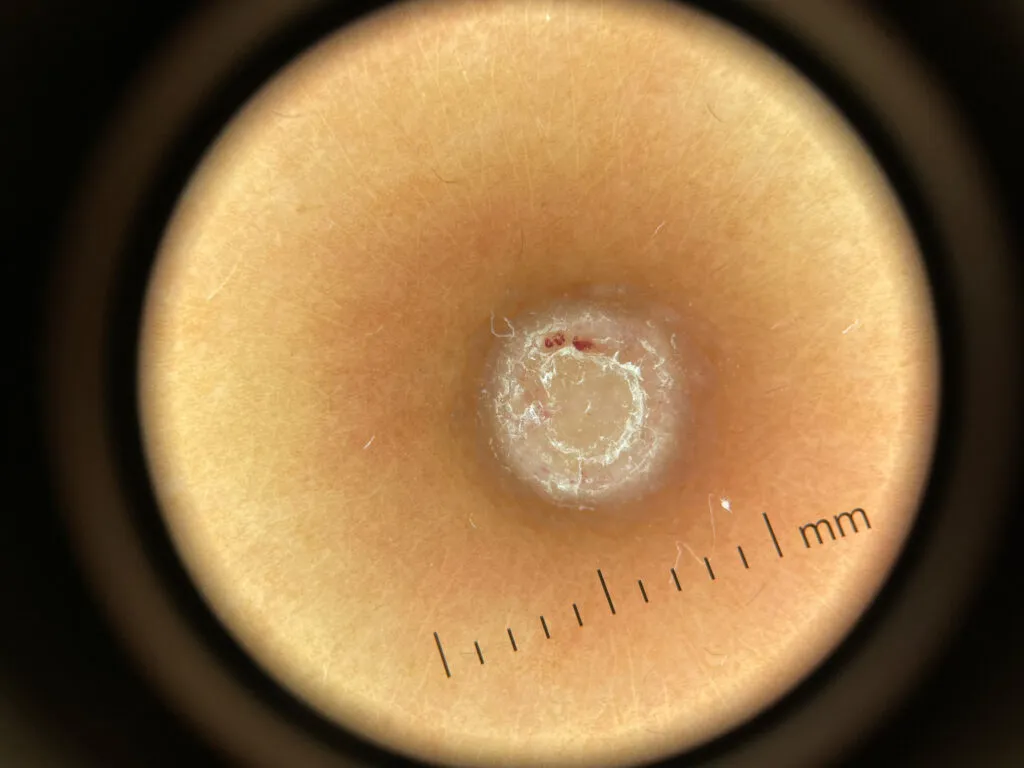
Persistent nevus under IBOOLO DE-3100

Dermoscopically, persistent nevi commonly show asymmetric but limited pigmentation patterns, with areas of light to dark brown color distributed irregularly over a whitish or pink scar background. The pigment network, when present, often appears broken, incomplete, or confined within the borders of the scar. Radial lines and globules may be seen, but they generally remain centrally located and do not extend beyond the scar’s margin. Fine linear or dotted vessels can also appear due to reactive vascular proliferation in the healing dermis. These characteristics contrast with recurrent melanoma, where pigmentation often extends beyond the scar edge, displays greater color variability including gray, blue, or black tones, and exhibits atypical network structures.

Persistent nevus
Persistent Nevus

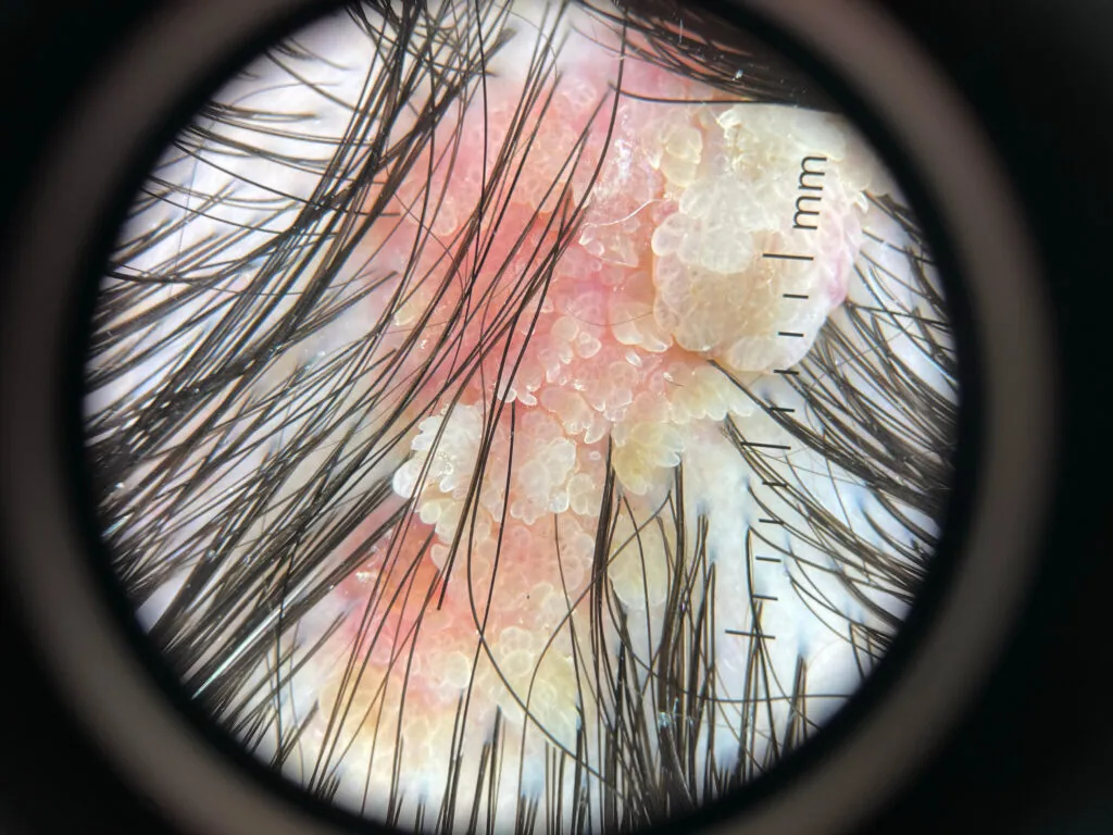
Seborrheic keratosis under IBOOLO DE-3100

The most consistent dermoscopic findings include multiple milia-like cysts, comedo-like openings, and fissures and ridges forming a brain-like or cerebriform surface pattern. The lesion surface often shows sharply demarcated borders with a waxy or keratotic appearance, correlating histologically with hyperkeratosis and acanthosis. Colors range from light brown to dark brown or black, depending on the thickness of the keratin and degree of pigmentation. In non-pigmented or lightly pigmented lesions, yellowish or whitish structureless areas may be observed. Some lesions show a network-like or reticular pattern, but this is usually pseudonetwork-like, caused by follicular openings rather than a true pigment network.

Seborrheic keratosis
Seborrheic Keratosis

Share this article

0

No products in the cart.

Ask

Have questions on gear or your order?

Our Gear Guides are here to help! Get personal advice from pro creatives

Name
Subject
Email address
How can we help?
Answers

Instant Answers

How can dermoscopy images be captured?

Dermoscopy images can be captured and stored in different ways, such as: • Using a smartphone or tablet with dermoscopic adapter, which consisted in the package.• Using a digital camera

Dermoscopy images can be captured and stored in different ways, such as:

• Using a smartphone or tablet with dermoscopic adapter, which consisted in the package.
• Using a digital camera with dermoscopic adapter, there’s 49mm screw size camera adapter available to order now.

Compatible phone/tablet models:
All iPhone models, 95% Android phones, 90% tablet. For phone/tablet size in 5.25-14mm

Compatible camera models:
All camera with built 49mm filter screw, such as Canon EOS 70D, 80D, 90D; Canon EOS R7, R10, R50, R100; Canon M100, M200, M50, Mark II; Canon G7X Mark III, Sony ZV-1

How can I connect my phone to my dermatoscope?

There’s universal phone adapter for all our dermoscopes. Please check the installation procedure bellow or watch operation guide. Smartphone Connector (1) Place phone adapter screw in the center of smartphone’s

There’s universal phone adapter for all our dermoscopes. Please check the installation procedure bellow or watch operation guide.

Smartphone Connector

(1) Place phone adapter screw in the center of smartphone’s main camera.
(2) Screw magnet attachment on phone adapter.
(3) Put dermoscope’s back ring and magnet attachment together

Take The Best Images

You need to adjust the focus ring after the dermoscpe connected on smartphone to get the best images.

How can I clean my dermoscopy after usage?

Cleaning your dermoscopy after usage is important to prevent cross-contamination and infection. The cleaning method may vary depending on the type and model of your dermoscopy, so you should always

Cleaning your dermoscopy after usage is important to prevent cross-contamination and infection. The cleaning method may vary depending on the type and model of your dermoscopy, so you should always follow the manufacturer’s instructions. However, some general steps are:

• Turn off and disconnect your dermoscopy from any power source or device.

• Wipe off any visible dirt or debris from the dermoscopy with a soft cloth or tissue.

• Disinfect the dermoscopy with an alcohol-based wipe or spray, or a disinfectant solution recommended by the manufacturer. Make sure to cover all surfaces, especially the lens and contact plate.

• Let the dermoscopy air dry completely before storing it in a clean and dry place.

• Do not use abrasive or corrosive cleaners, solvents, or detergents that may damage the dermoscopy.

• Do not immerse the dermoscopy in water or any liquid, unless it is waterproof and designed for immersion.

You should clean your dermoscopy after each use, or at least once a day if you use it frequently. You should also check your dermoscopy regularly for any signs of damage or malfunction, and contact the manufacturer or service provider if needed.

Polarized VS Non-polarized Dermoscopy

A dermoscopy is a device that allows the examination of skin lesions with magnificationand illumination. By revealing subsurface structures and patterns that are not visible tothe naked eye. It can

A dermoscopy is a device that allows the examination of skin lesions with magnificationand illumination. By revealing subsurface structures and patterns that are not visible tothe naked eye. It can improve the diagnose accuracy of skin lesions, such as melanoma,basal cell carcinoma, seborrheic keratosis, etc.

There are two main types of dermoscopy: Non polarized and polarized dermoscopy.We’ve fitted most of our dermoscopys with polarized and non-polarized light. They canbe used in multiple skin structures.

Non-polarized contact Mode

In non-polarized mode, the instrument can provide information about the superficialskin structures, such as milia-like cysts, comedo-like openings, and pigment in theepidemis.

The dermoscopy requires applying a liquid such as mineral oil or alcohol to the skin andplacing the lens in contact with the skin. This reduces surface reflection and enhancesthe view of subsurface structures.

Image with non-polarized light (DE-3100)

Polarized contact Mode

In polarized mode, the instrument allows for visualization for deeper skin structures,such as blood vessels, collagen, and pigment in the dermis.

The dermoscopy does not need to be in contact with the skin or use any liquid. Theirpolarized light can help to eliminate surface reflection and allow visualization ofvascular structures.

Image with polarized light (DE-3100)

Polarized non-contact Mode

The dermoscopy can also use polarized light to examine the skin without direct contact.

In polarized non-contact mode, the instrument allows for examination infected areasand lesions that are painful for the patient, or the difficult to contact pigmented lesions,such as nails and narrow areas.

The contact plate should be removed in this mode, and it does not require applying aliquid to the skin. As it doesn’t require pressure or fluid application on the skin, it canalso avoid cross-contamination and infection risk.

Image in polarized non-contact mode (DE-3100)

How effectiveness is dermoscopy

Compared with visual inspection, the dermoscopy can be used to capture and store skin lesion photos, which play an important role in early skin cancer examination. The dermoscopy allows the

Compared with visual inspection, the dermoscopy can be used to capture and store skin lesion photos, which play an important role in early skin cancer examination.

The dermoscopy allows the examination of skin lesions with magnification and illumination. This can be greatly avoiding the factors that cause interference to visual detection. Such as lighting, skin color, hair and cosmetics.

Several studies have demonstrated that dermoscopy is useful in the identification of melanoma, when used by a trained professional.

It may improve the accuracy of clinical diagnosis by up to 35%
It may reduce the number of harmless lesions that are removed
In primary care, it may increase the referral of more worrisome lesions and reduce the referral of more trivial ones

A 2018 Cochrane meta-analysis published the accuracy of dermoscopy in the detection.

Table 1. Accuracy of dermoscopy in the detection of melanoma in adults
Detection Method Sensitivity, % Specificity, % Positive Likelihood Ratio NegativeLikelihood Ratio
Visual inspection alone (in person) 76 75 3.04 0.32
Dermoscopy with visual inspection (in person) 92 95 18 0.08
Image-based visual inspection alone (not in person) 47 42 0.81 1.3
Dermoscopy with image-based visual inspection (not in person) 81 82 4.5 0.23
ROC—receiver operating characteristic. *Estimated sensitivity calculated on the summary ROC curve at a fixed specificity of 80%.

As we can see, the dermoscope can improve the accuracy of diagnosis of skin lesions, especially melanoma.

Table 1. Accuracy of dermoscopy in the detection of melanoma in adults
Detection Method Sensitivity, % Specificity, % Positive Likelihood Ratio NegativeLikelihood Ratio
Visual inspection alone (in person) 79 77 3.4 0.27
Dermoscopy with visual inspection (in person) 93 99 93 0.07
Image-based visual inspection alone (not in person) 85 87 6.5 0.17
Dermoscopy with image-based visual inspection (not in person) 93 96 23 0.07
ROC—receiver operating characteristic. *Estimated sensitivity calculated on the summary ROC curve at a fixed specificity of 80%.

Characteristics of the dermatoscopic structure of the skin lesions include:

• Symmetry or asymmetry
• Homogeny/uniformity (sameness) or heterogeny (structural differences across the lesion)
• Distribution of pigment: brown lines, dots, clods and structureless areas
• Skin surface keratin: small white cysts, crypts, fissures
• Vascular morphology and pattern: regular or irregular
• Border of the lesion: fading, sharply cut off or radial streaks
• Presence of ulceration

There are specific dermoscopic patterns that aid in the diagnosis of the following pigmented skin lesions:

• Melanoma
• Moles (benign melanocytic naevus)
• Freckles (lentigos)
• Atypical naevi
• Blue naevi
• Seborrhoeic keratosis
• Pigmented basal cell carcinoma
• Haemangioma

We use cookies on this website to provide a better user experience. By continuing to browse the website, you are giving your consent to receive cookies on this site. For more details please read our Privacy Policy.

Hot Search Terms