Skin Lesions Under IBOOLO DE-4100 Dermoscope – IBOOLO

Shenzhen Iboolo Optics Co.Ltd has been manufacturing Macro lens, Woods Lamp, Dermatoscope and Microscope ever since the company was established in 2012. Our company offers a wide variety of goods that can be tailored to meet your specific requirements.

Article

Skin Lesions Under IBOOLO DE-4100 Dermoscope

This article mainly aims to share a series of lesion images captured by users with the IBOOLO DE-4100 dermatoscope. The IBOOLO DE-4100 offers four lighting modes: polarized light, non-polarized light, amber light, and polarized-amber mixed light. The images presented in this article were primarily taken using the polarized mode. Basal Cell Carcinoma under IBOOLO DE-4100  On…

This article mainly aims to share a series of lesion images captured by users with the IBOOLO DE-4100 dermatoscope. The IBOOLO DE-4100 offers four lighting modes: polarized light, non-polarized light, amber light, and polarized-amber mixed light. The images presented in this article were primarily taken using the polarized mode.

Basal Cell Carcinoma under IBOOLO DE-4100 

On dermoscopy, basal cell carcinoma (BCC) is characterized by the absence of a pigment network and the presence of specific structures such as arborizing telangiectasias, blue-gray ovoid nests, maple-leaf–like areas, spoke-wheel areas, and ulceration. Polarized light can reveal shiny white structures, and in superficial BCC, short fine telangiectasias and multiple small erosions are common.

Basal Cell Carcinoma IBOOLO
Basal Cell Carcinoma

Melanoma under IBOOLO DE-4100 

Melanoma under dermoscopy typically displays asymmetry and an atypical pigment network, often accompanied by irregular dots and globules, streaks or pseudopods, and the presence of a negative network. Additional features include blue-white veil, regression structures such as peppering and scar-like depigmentation, shiny white structures, and polymorphous or atypical vessels. These features, particularly shiny white structures, pseudopods, irregular pigmentation, blue-white veil, and peppering, are strongly associated with melanoma diagnosis.

Melanoma IBOOLO
Melanoma

Actinic Keratosis under IBOOLO DE-4100

Actinic keratosis exhibits the so-called strawberry pattern: a pink-to-red background surmounted by a white-yellow surface scale arranged in a geometric, tile-like fashion. Under polarised light, fine wavy hairpin vessels are often visible, and rosettes—four white dots arranged in a square—may be seen. The scale is adherent and cannot be removed without trauma, distinguishing the lesion from seborrhoeic keratosis.

Actinic Keratosis IBOOLO
Actinic Keratosis

Cherry Angioma under IBOOLO DE-4100

Cherry angioma, a common type of acquired hemangioma, shows a distinctive lacunar pattern under dermoscopy. This consists of well-demarcated, round to oval, red, maroon, or blue-black lacunae separated by fibrous septa. Thrombosed areas may appear darker, and the lacunar arrangement is considered diagnostic.

Cherry angioma IBOOLO
Cherry Angioma

Spider Angioma under IBOOLO DE-4100

Spider angioma, also known as spider telangiectasis, is characterized by a central red arteriole with radially arranged thin capillaries. On dermoscopy, the blanch-and-refill sign is diagnostic: pressure applied to the central vessel causes blanching of the entire lesion, with rapid refill upon release. This pattern reflects the vascular structure of the lesion.

Spider angioma IBOOLO
Spider Angioma

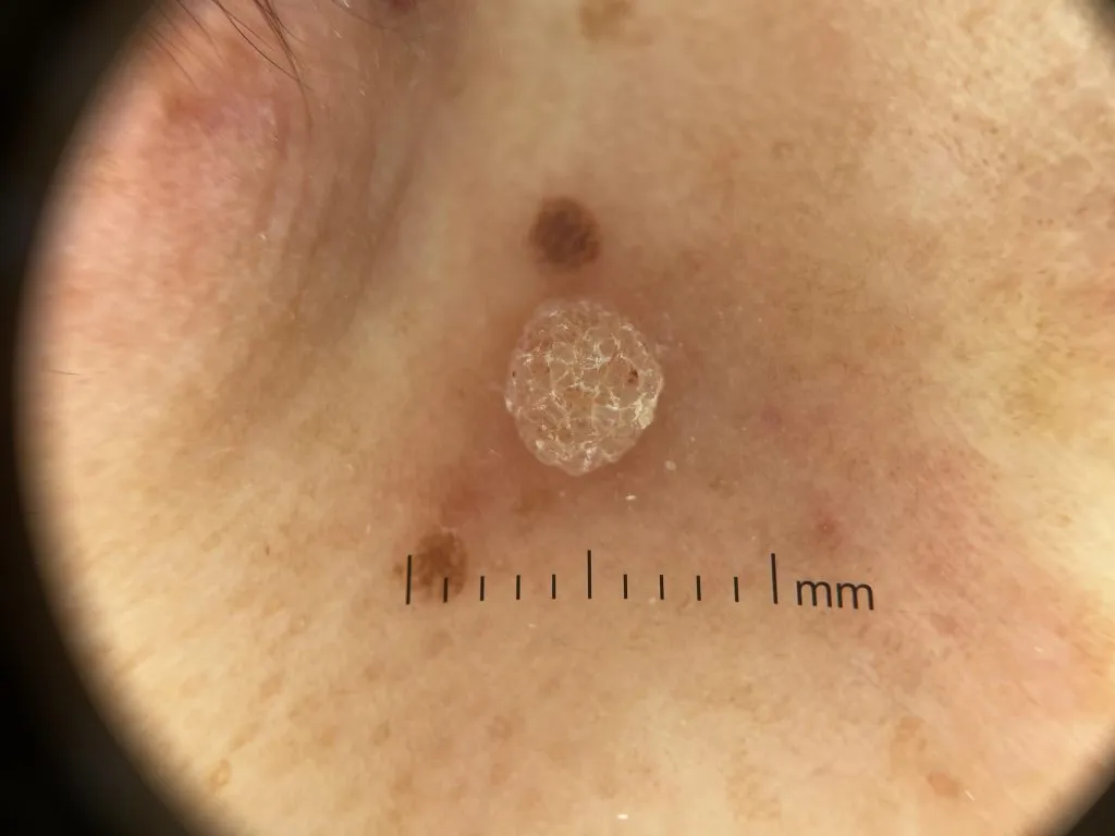
Common Wart under IBOOLO DE-4100

Common wart shows a papillomatous, hyperkeratotic surface on dermoscopy, often with multiple densely packed papillae. Each papilla contains a central red or black dot or loop corresponding to thrombosed capillaries. The skin lines are interrupted, and plantar warts frequently display numerous hemorrhagic dots within a yellowish papilliform surface, which helps differentiate them from calluses or corns.

Common wart IBOOLO
Common Wart

Blue Nevus under IBOOLO DE-4100

A blue nevus under dermoscopy typically shows uniform, structureless pigmentation that appears steel-blue, blue-gray, blue-brown, or blue-black; this homogeneous blue appearance is the hallmark feature, and in some cases small dots or globules may also be observed or rare depigmented scarlike areas peripherally.

blue nevus IBOOLO
Blue Nevus

Sebaceous Nevus under IBOOLO DE-4100

A sebaceous nevus during dermoscopic examination frequently reveals aggregated yellowish or brown globules and lobular structures on a yellow background. In childhood, these features appear as clustered lobules in yellow background; verrucous or warty stages show whitish-yellow lobular or grayish papillary appearance, while nodular stages may display homogeneous yellow–whitish pigmentation.

sebaceous nevus IBOOLO
Sebaceous Nevus

Verrucous Epidermal Nevus under IBOOLO DE-4100

A verrucous epidermal nevus demonstrates a mix of distinctive dermoscopic structures. Common findings include large brown circles (oval or round structures with darker brown edges surrounding lighter areas), thick branched brown lines, brown dots and globules arranged in lines or serpiginous patterns, exophytic white and brown papillary structures, cerebriform or cobblestone patterns, thick adherent scales, and dotted vessels; these features may overlap with seborrheic keratosis, but the presence of cerebriform pattern and large brown circles may be particularly specific to this nevus.

verrucous epidermal nevus IBOOLO
Verrucous Epidermal Nevus

Seborrheic Keratosis under IBOOLO DE-4100

Seborrheic keratosis exhibits multiple, well-characterized dermoscopic signs. These include milia-like cysts, comedo-like (pseudohorn) openings, fissures and ridges often describing a cerebriform or “gyri and sulci” pattern, moth-eaten or sharply demarcated borders, fingerprint-like structures, “fat-finger” projections, and hairpin (looped) vessels often surrounded by a whitish halo.

Seborrheic keratosis IBOOLO
Seborrheic Keratosis

Acral Junctional Nevu under IBOOLO DE-4100

An acral junctional nevus—interpreted as a benign melanocytic nevus arising at the border of acral skin such as the sole—displays under dermoscopy patterns typical of benign acral melanocytic nevi. These include the parallel furrow pattern (pigmentation following the skin sulci), lattice-like pattern (pigmentation along furrows with crossing linear bands), fibrillar pattern (fine streaks crossing dermatoglyphic ridges, often due to pressure), homogeneous pattern, or globular variants; among these, parallel furrow is the prototypical sign of benignity, and fibrillar and lattice-like variants may also appear, sometimes mixed within the same lesion.

acral junctional nevus IBOOLO
Acral Junctional Nevus

Café-au-lait Macule under IBOOLO DE-4100

A café-au-lait macule under dermoscopy shows a uniform light-brown background with a faint but consistently reticulated brown pigment network; in early lesions, this may appear as focal thickening of the network forming tiny arcuate lines, while in darker or more mature lesions the pigment network becomes uniformly thickened and more evident. These lesions lack structures such as dots, globules, streaks, or vascular patterns that are typically seen in melanocytic or vascular conditions, distinguishing café-au-lait macules from nevi or melanomas.

café-au-lait macule IBOOLO
Café-au-lait Macule

Share this article

0

No products in the cart.

Ask

Have questions on gear or your order?

Our Gear Guides are here to help! Get personal advice from pro creatives

Name
Subject
Email address
How can we help?
Answers

Instant Answers

How can dermoscopy images be captured?

Dermoscopy images can be captured and stored in different ways, such as: • Using a smartphone or tablet with dermoscopic adapter, which consisted in the package.• Using a digital camera

Dermoscopy images can be captured and stored in different ways, such as:

• Using a smartphone or tablet with dermoscopic adapter, which consisted in the package.
• Using a digital camera with dermoscopic adapter, there’s 49mm screw size camera adapter available to order now.

Compatible phone/tablet models:
All iPhone models, 95% Android phones, 90% tablet. For phone/tablet size in 5.25-14mm

Compatible camera models:
All camera with built 49mm filter screw, such as Canon EOS 70D, 80D, 90D; Canon EOS R7, R10, R50, R100; Canon M100, M200, M50, Mark II; Canon G7X Mark III, Sony ZV-1

How can I connect my phone to my dermatoscope?

There’s universal phone adapter for all our dermoscopes. Please check the installation procedure bellow or watch operation guide. Smartphone Connector (1) Place phone adapter screw in the center of smartphone’s

There’s universal phone adapter for all our dermoscopes. Please check the installation procedure bellow or watch operation guide.

Smartphone Connector

(1) Place phone adapter screw in the center of smartphone’s main camera.
(2) Screw magnet attachment on phone adapter.
(3) Put dermoscope’s back ring and magnet attachment together

Take The Best Images

You need to adjust the focus ring after the dermoscpe connected on smartphone to get the best images.

How can I clean my dermoscopy after usage?

Cleaning your dermoscopy after usage is important to prevent cross-contamination and infection. The cleaning method may vary depending on the type and model of your dermoscopy, so you should always

Cleaning your dermoscopy after usage is important to prevent cross-contamination and infection. The cleaning method may vary depending on the type and model of your dermoscopy, so you should always follow the manufacturer’s instructions. However, some general steps are:

• Turn off and disconnect your dermoscopy from any power source or device.

• Wipe off any visible dirt or debris from the dermoscopy with a soft cloth or tissue.

• Disinfect the dermoscopy with an alcohol-based wipe or spray, or a disinfectant solution recommended by the manufacturer. Make sure to cover all surfaces, especially the lens and contact plate.

• Let the dermoscopy air dry completely before storing it in a clean and dry place.

• Do not use abrasive or corrosive cleaners, solvents, or detergents that may damage the dermoscopy.

• Do not immerse the dermoscopy in water or any liquid, unless it is waterproof and designed for immersion.

You should clean your dermoscopy after each use, or at least once a day if you use it frequently. You should also check your dermoscopy regularly for any signs of damage or malfunction, and contact the manufacturer or service provider if needed.

Polarized VS Non-polarized Dermoscopy

A dermoscopy is a device that allows the examination of skin lesions with magnificationand illumination. By revealing subsurface structures and patterns that are not visible tothe naked eye. It can

A dermoscopy is a device that allows the examination of skin lesions with magnificationand illumination. By revealing subsurface structures and patterns that are not visible tothe naked eye. It can improve the diagnose accuracy of skin lesions, such as melanoma,basal cell carcinoma, seborrheic keratosis, etc.

There are two main types of dermoscopy: Non polarized and polarized dermoscopy.We’ve fitted most of our dermoscopys with polarized and non-polarized light. They canbe used in multiple skin structures.

Non-polarized contact Mode

In non-polarized mode, the instrument can provide information about the superficialskin structures, such as milia-like cysts, comedo-like openings, and pigment in theepidemis.

The dermoscopy requires applying a liquid such as mineral oil or alcohol to the skin andplacing the lens in contact with the skin. This reduces surface reflection and enhancesthe view of subsurface structures.

Image with non-polarized light (DE-3100)

Polarized contact Mode

In polarized mode, the instrument allows for visualization for deeper skin structures,such as blood vessels, collagen, and pigment in the dermis.

The dermoscopy does not need to be in contact with the skin or use any liquid. Theirpolarized light can help to eliminate surface reflection and allow visualization ofvascular structures.

Image with polarized light (DE-3100)

Polarized non-contact Mode

The dermoscopy can also use polarized light to examine the skin without direct contact.

In polarized non-contact mode, the instrument allows for examination infected areasand lesions that are painful for the patient, or the difficult to contact pigmented lesions,such as nails and narrow areas.

The contact plate should be removed in this mode, and it does not require applying aliquid to the skin. As it doesn’t require pressure or fluid application on the skin, it canalso avoid cross-contamination and infection risk.

Image in polarized non-contact mode (DE-3100)

How effectiveness is dermoscopy

Compared with visual inspection, the dermoscopy can be used to capture and store skin lesion photos, which play an important role in early skin cancer examination. The dermoscopy allows the

Compared with visual inspection, the dermoscopy can be used to capture and store skin lesion photos, which play an important role in early skin cancer examination.

The dermoscopy allows the examination of skin lesions with magnification and illumination. This can be greatly avoiding the factors that cause interference to visual detection. Such as lighting, skin color, hair and cosmetics.

Several studies have demonstrated that dermoscopy is useful in the identification of melanoma, when used by a trained professional.

It may improve the accuracy of clinical diagnosis by up to 35%
It may reduce the number of harmless lesions that are removed
In primary care, it may increase the referral of more worrisome lesions and reduce the referral of more trivial ones

A 2018 Cochrane meta-analysis published the accuracy of dermoscopy in the detection.

Table 1. Accuracy of dermoscopy in the detection of melanoma in adults
Detection Method Sensitivity, % Specificity, % Positive Likelihood Ratio NegativeLikelihood Ratio
Visual inspection alone (in person) 76 75 3.04 0.32
Dermoscopy with visual inspection (in person) 92 95 18 0.08
Image-based visual inspection alone (not in person) 47 42 0.81 1.3
Dermoscopy with image-based visual inspection (not in person) 81 82 4.5 0.23
ROC—receiver operating characteristic. *Estimated sensitivity calculated on the summary ROC curve at a fixed specificity of 80%.

As we can see, the dermoscope can improve the accuracy of diagnosis of skin lesions, especially melanoma.

Table 1. Accuracy of dermoscopy in the detection of melanoma in adults
Detection Method Sensitivity, % Specificity, % Positive Likelihood Ratio NegativeLikelihood Ratio
Visual inspection alone (in person) 79 77 3.4 0.27
Dermoscopy with visual inspection (in person) 93 99 93 0.07
Image-based visual inspection alone (not in person) 85 87 6.5 0.17
Dermoscopy with image-based visual inspection (not in person) 93 96 23 0.07
ROC—receiver operating characteristic. *Estimated sensitivity calculated on the summary ROC curve at a fixed specificity of 80%.

Characteristics of the dermatoscopic structure of the skin lesions include:

• Symmetry or asymmetry
• Homogeny/uniformity (sameness) or heterogeny (structural differences across the lesion)
• Distribution of pigment: brown lines, dots, clods and structureless areas
• Skin surface keratin: small white cysts, crypts, fissures
• Vascular morphology and pattern: regular or irregular
• Border of the lesion: fading, sharply cut off or radial streaks
• Presence of ulceration

There are specific dermoscopic patterns that aid in the diagnosis of the following pigmented skin lesions:

• Melanoma
• Moles (benign melanocytic naevus)
• Freckles (lentigos)
• Atypical naevi
• Blue naevi
• Seborrhoeic keratosis
• Pigmented basal cell carcinoma
• Haemangioma

We use cookies on this website to provide a better user experience. By continuing to browse the website, you are giving your consent to receive cookies on this site. For more details please read our Privacy Policy.

Hot Search Terms