


High-Performance 365nm UV Lamp Technology for Medical and Professional Diagnostics
In the specialized field of optical diagnostics, the precision of the light source determines the accuracy of the results. The 365nm UV lamp, specifically within the UVA spectrum, has become the gold standard for healthcare professionals and industrial inspectors alike. Unlike standard ultraviolet lights, a true 365nm wavelength provides the necessary excitation energy to induce fluorescence in various biological and chemical substances without the interference of visible light or the hazards associated with shorter UVB or UVC rays. IBOOLO’s DE-215 represents the pinnacle of this technology, engineered to meet the rigorous demands of clinical environments.
The Science Behind 365nm UV Wavelength Accuracy
The effectiveness of a UV lamp is defined by its peak wavelength and spectral purity. Many consumer-grade ultraviolet lights operate at 395nm, which emits a significant amount of visible purple light that can mask subtle fluorescence. A professional 365nm UV lamp, however, produces a much "cleaner" light that targets the specific absorption peaks of fluorophores found in skin, fungi, and specialized industrial dyes.
Why Medical Professionals Require 365nm Precision
In clinical settings, the 365nm UV lamp is primarily utilized as a Wood’s Lamp. This specific wavelength is essential because it penetrates the epidermal layer effectively, allowing clinicians to visualize abnormalities that are invisible under standard ambient lighting. By maintaining a stable radiation intensity of 3.5 mW/cm2, IBOOLO ensures that the fluorescence remains consistent throughout the examination, providing reliable data for longitudinal patient tracking.
Primary Applications in Clinical Dermatology
A 365nm UV lamp is an indispensable diagnostic tool for the modern dermatology clinic. Its ability to reveal skin irregularities and microorganisms through fluorescence makes it a first-line instrument for several critical assessments.
Identification of Pigmentary Disorders
Conditions such as vitiligo and melasma require precise visualization of pigment loss or gain. Under a 365nm UV lamp, depigmented areas in vitiligo appear as bright, chalk-white patches with sharp borders. This contrast allows dermatologists to define the exact extent of the condition, which is often difficult to discern on fair-skinned patients under white light.
Detection of Bacterial and Fungal Infections
Certain microorganisms produce characteristic fluorescence when exposed to 365nm UV light. For example, Erythrasma exhibits a coral-red glow due to porphyrin production by Corynebacterium minutissimum. Similarly, specific fungal infections like Tinea Capitis will fluoresce brilliant blue-green. The immediate visual feedback provided by the lamp allows for rapid triage and treatment planning.
Technical Superiority: The DE-215 Wood’s Lamp Engineering
Choosing the right 365nm UV lamp involves looking beyond the light bulb. The engineering of the device determines its longevity, safety, and clinical utility.
- Enhanced Optical Field of View: The DE-215 features a 50mm diameter lens with 4.5X magnification, allowing for high-resolution inspection of skin lesions while maintaining the necessary distance for safety and comfort.
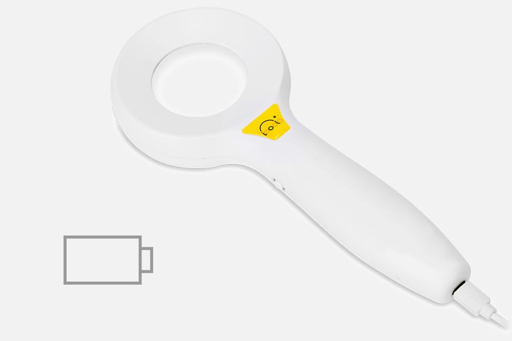
- Extended Battery Performance: Equipped with a 2000mAh lithium-ion battery, this professional lamp supports up to 6 hours of continuous operation, ensuring it is always ready for a full day of patient consultations.
- Uniform Radiation Distribution: Unlike cheap LED alternatives that produce hot spots, our optical system delivers uniform 365nm radiation across the entire field of view, preventing false negatives during diagnosis.
- Ergonomic and Durable Design: The lightweight construction is optimized for ease of use, while the USB-C charging interface ensures compatibility with modern clinical infrastructure.
Safety Standards and Regulatory Compliance
When implementing a 365nm UV lamp in a professional setting, adherence to safety protocols is paramount. While UVA is the safest part of the UV spectrum, high-intensity devices must be used with care. IBOOLO devices are designed with these safety parameters in mind, ensuring that radiation levels are optimized for diagnostic efficacy without excessive exposure risk.
Our commitment to quality is reflected in our ISO 13485 certification and FDA registration. For hospitals and medical distributors, this means every 365nm UV lamp we produce meets the international standards for medical device safety and performance. We provide comprehensive spectral data to prove that our lamps emit at the peak 365nm mark, minimizing harmful UVB/UVC leakage.
Comparison: 365nm vs. 395nm UV Lamps
| Feature | 365nm UV Lamp (Professional) | 395nm UV Lamp (Consumer) |
|---|---|---|
| Fluorescence Visibility | High (Minimal visible light interference) | Low (Heavy purple light masking) |
| Diagnostic Accuracy | Clinical Grade | General Purpose Only |
| Primary Use | Medical, Forensics, NDT | Pet Stains, Resin Curing |
| Contrast Ratio | Superior | Poor |
Frequently Asked Questions About 365nm UV Lamps
What is the difference between a Wood's Lamp and a 365nm UV Lamp?
A Wood’s Lamp is essentially a specialized 365nm UV lamp used for medical diagnostics. The term refers to the clinical application, while 365nm refers to the specific wavelength of light used to excite fluorescence in biological samples.
Can a 365nm UV lamp be used for teledermatology?
Yes. When combined with high-resolution photography, the images captured under a 365nm UV lamp provide invaluable data for remote consultations, particularly for tracking the progression of vitiligo or acne treatments.
Does the DE-215 require specialized training?
While the device is user-friendly, interpreting the results of a 365nm UV lamp examination requires knowledge of dermatoscopy and skin fluorescence patterns. We provide a detailed user manual to assist professionals in optimizing their diagnostic workflow.
Recommended reading
365nm UV Lamp for enhanced visualization in dermatology - IBOOLO
IBOOLO 12W 365nm UV lamp delivers optimized illumination for diagnosing bacterial, and fungal infections and skin pigment disorders and enhances visualization of skin conditions like tinea capitis.
Dermoscopy of Amelanotic Melanoma – IBOOLO
Melanoma is the most invasive and dangerous of the common forms of skin cancer with the highest risk of death. Melanoma moves very quickly, it can spread to other parts of the body. If untreated, melanoma can became life-threatening even in 6 weeks. Amelanotic melanoma is the highly aggressive form of melanoma that does not...
China Customized Dermatoscopio Digital Suppliers & Wholesalers Focused on High-End Products - IBOOLO
Through advanced China production techniques, our suppliers & wholesalers facilities supply customized dermatoscopio digitals merging durable frames and peerless optics for precise magnification.
Dermatology UV 365nm DE-215 Woods Lamp
Dermatology UV 365nm DE-215 Woods Lamp


- 4.5 x Magnification
- 60mm Lens Diameter
- 20 LEDs (15 White, 5 UV 365nm)
- 2 Types Colour Lighting
- Automatic shutdown
- Ultra long life battery
| Material | Plastic & Glass |
| Optical Design | All glass, 1 element 1 group, anti-reflection coating |
| Lens Diameter | 50mm(front); 50mm(rear) |
| Magnification | 4.5x |
| Distortion | 5% |
| Ultraviolet Wave Length | 5 LEDs 365nm UV, 15 while LEDs |
| Resolution | 300 LP/MM (Axis) 250 LP/MM (Edges) |
| LED Type | SMD LED beads |
| Battery Capacity | 2000mAh Lithium ion |
| Charging | USB-C |
| Working Time | 6-8 hours |
| Focal Length | 15-30cm |
| Dimension | Φ100mm*H34mm*L240mm |
| Weight | 162g |


Related documents & accessories

DE-3100 PRO Dermatoscope
$599.00

DE-300 Dermatoscope
$109.00
What It Has
- 365nm UV Light:A Wood’s lamp has broadband light sources that emit light at wavelengths between 320nm and 400nm, with a peak at 365nm.
- 4.5X Magnification:Low distortion magnify
- 2000mAh Battery: Offering up to 6 hours long time and stable diagnosis.
In The Box
- DE-215 Woods Lamp
- USB Type-C Charging Cable
- Microfiber Cloth
- User Manual
Specs
- Lens Diameter:50mm
- Magnification:4.5X
- Ultraviolet Wavelength: 365nm
- Radiation Intensity: 3.5 mW/cm2
- Battery Capacity:2000mAh
- Charging:USBType-C
- Working Time:2-6 hours
- Dimension:240mm*100mm*34mm(L*W*H)
Clinical Applications
The Wood's lamp is used to identify the extent of pigmented or depigmented patches and to detect fluorescence. Normal healthy skin is slightly blue but shows white spots where there is thickened skin, yellow where it is oily, and purple spots where it is dehydrated. Clothing lint often shines bright white.

Vitiligo

Fluorescence

Tinea Capitis

Fungal Inflection
What Makes it Unique
Woods lamps use ultraviolet light to reveal skin abnormalities that can’t be seen with human eyes. Build with 60mm field view and no cross-infection design, this Woods lamp can be held about 10-30 cm away from the skin for detection. The examination is painless and safe.
Why Choose IBOOLO DE-215 Woods Lamp
High Performance

High Performance
To effectively reveal skin abnormalities, extensive and uniform radiation light are needed.
Practical
The 60mm diameter design make sure there’s no unnecessary corners and gaps to catch hair or scales in.
Practical

Durable
Durable
Thanks to the solid body design and long life battery (2000mAh), the lamp offers long time and stable diagnosis.
You might also like

DE-4100 Dermatoscope
$699.00

DE-3100 Dermatoscope
$499.00

DE-400 Dermatoscope
$179.00
DE-300 Dermatoscope
$109.00

Reviews
1 review for DE-215 Woods Lamp
Only logged in customers who have purchased this product may leave a review.
Learn More

iPhone 17 Pro Max Dermoscopy: How to Optimize High-Resolution Skin Imaging with IBOOLO Universal Adapters
The release of the iPhone 17 Pro Max marks a significant leap in mobile imaging technology. With its advanced sensor upgrades and computational photography, it offers unprecedented potential for clinical…

What Are the Differences Between the DE-3100 and DE-4100, and the Pro version?
A dermatoscope is more than just a magnifying glass with a light. It utilizes polarized and non-polarized light to eliminate surface reflection from the stratum corneum, allowing a clear view…

What Are the Differences Between IBOOLO DE-300, DE-400, and DE-500?
A dermatoscope is a medical optical device designed to visualize skin structures that are not visible to the naked eye. By combining magnification with controlled illumination, a dermatoscope allows clearer…


Kim Long United States
–
United States
–
Excellent product! The Wood’s lamp is incredibly effective and easy to use. It provides clear, bright UV light.