-
×
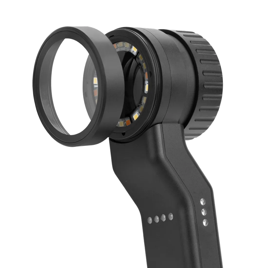
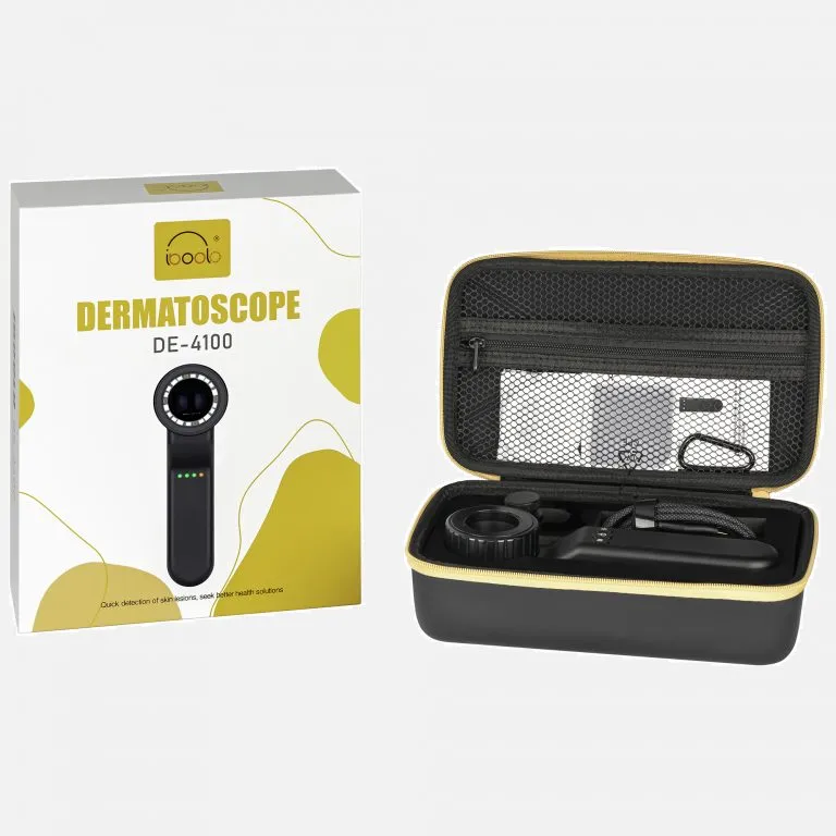
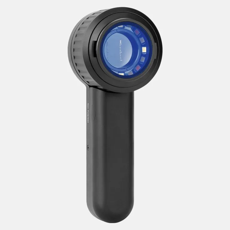
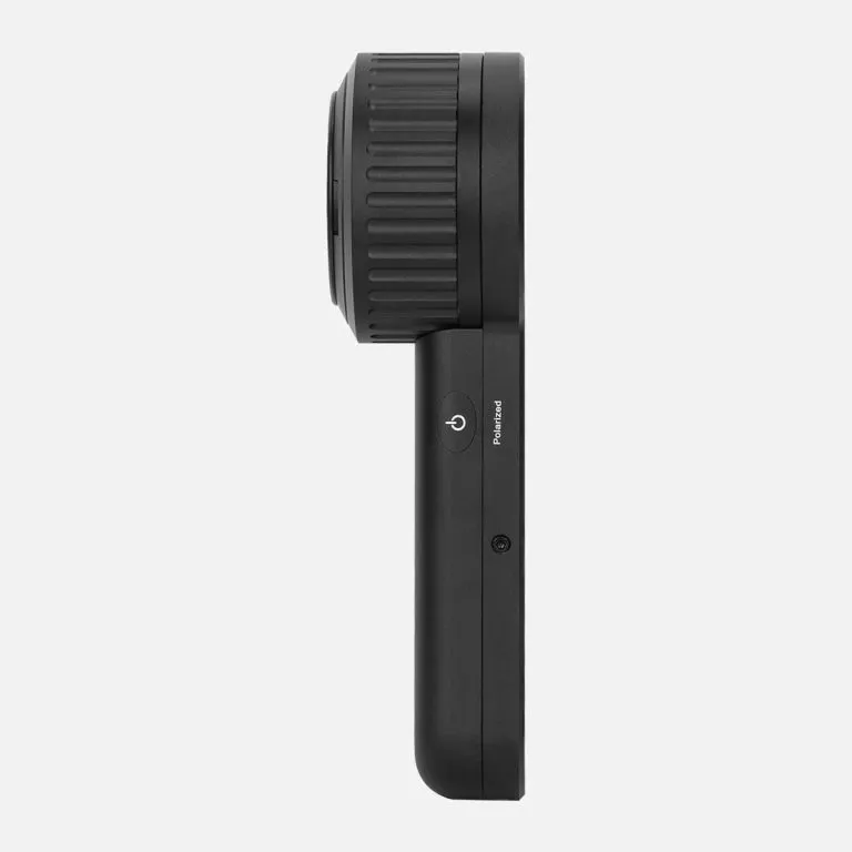
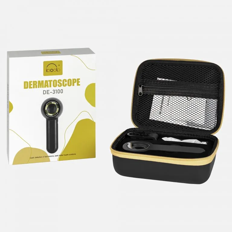
 DE-500 Dermatoscope
1 × $399.00
DE-500 Dermatoscope
1 × $399.00
Subtotal: $399.00
Message reply within 1 day
Free national shipping
100% satisfaction
2 years warranty
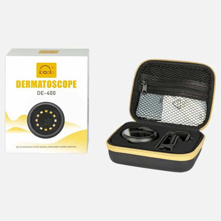
As one of the best reliable dermatoscope suppliers globally,
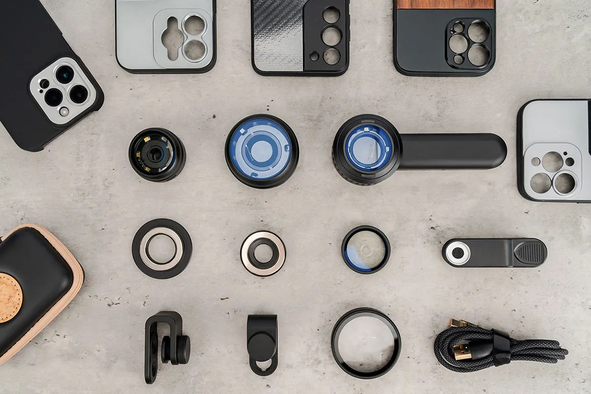
Iboolo offers dermatoscopes, woods lamps, phone adapters, & so much more.
Get world-leading product quality at an affordable price, with 24/7 support for a quotation.
Buy professional dermatoscopes online. Explore our digital dermoscopic cameras & electronic systems. FDA registered, ISO 13485 quality. Best dermatoscope price for clinics.
Looking for the best dermatoscopes for sale? IBOOLO provides clinical-grade electronic dermatoscope and digital dermatoscope systems for dermatologists and medical practitioners. As an ISO 13485 certified manufacturer, we offer high-resolution optics with FDA registration, ensuring you get premium diagnostic tools at a competitive dermoscopy price.
Understanding the dermatoscope cost is essential for budgeting your practice. Below is a comparison of our professional-grade devices to help you find the best dermascope price for your needs.
| Model Category | Key Features | Best For | Estimated Price |
|---|---|---|---|
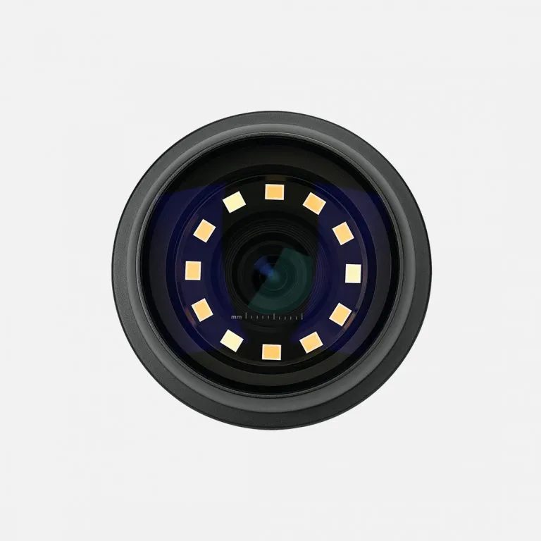
| Electronic Dermatoscope | HD Polarized Light, 30x Zoom | Professional Clinical Diagnosis | Check Best Price |
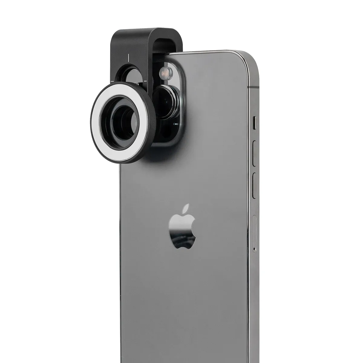
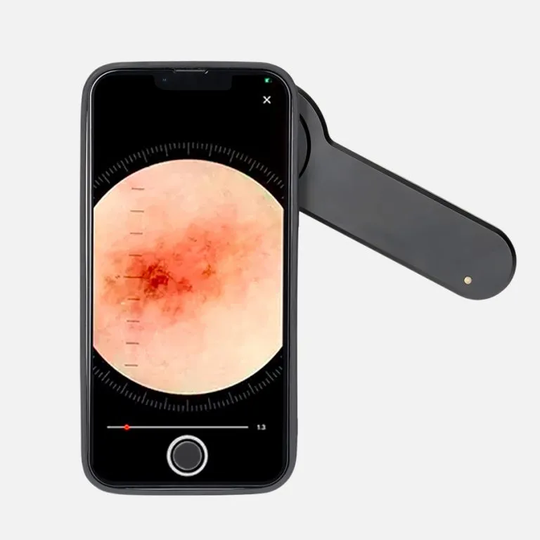
| Digital Dermatoscope | Smartphone Sync, Video Capture | Telemedicine & Documentation | Check Best Price |
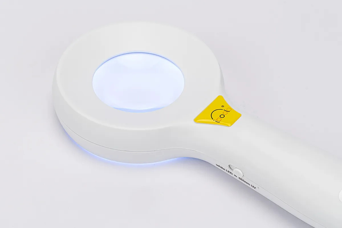
| Dermatoscopio Digital | Portable, Multi-spectral LED | Mobile Skin Screening | Check Best Price |
| Cheap Dermatoscope (Entry) | Professional Optics, 10x Fixed | Students & General Practice | Affordable Pricing |
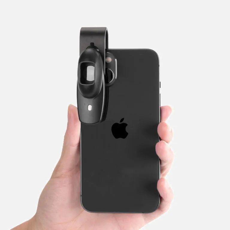
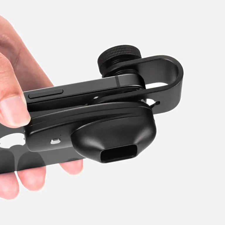
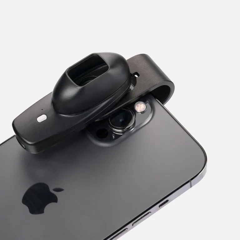
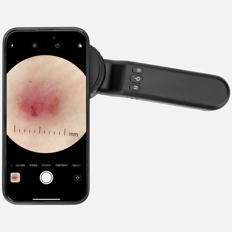
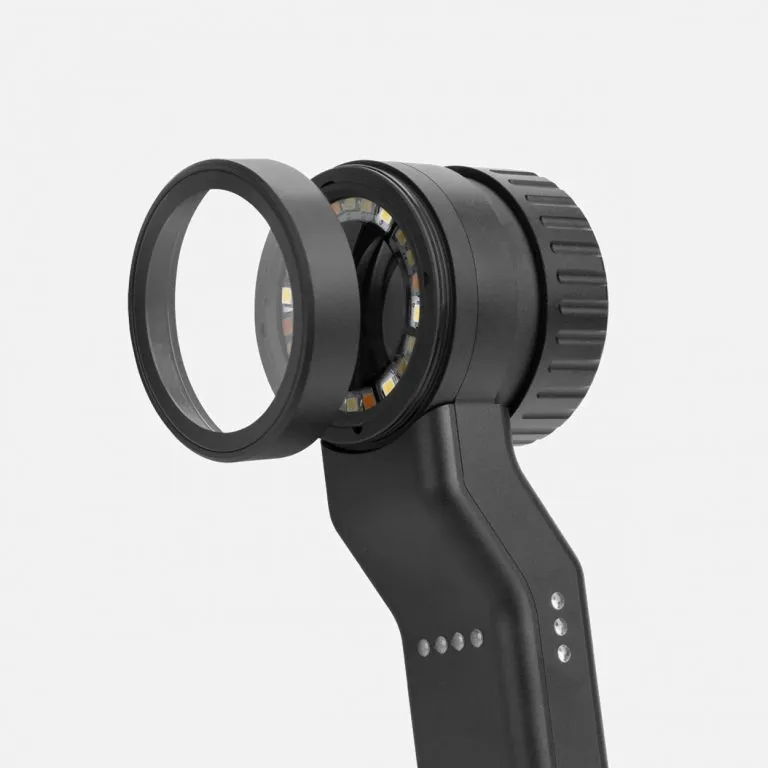
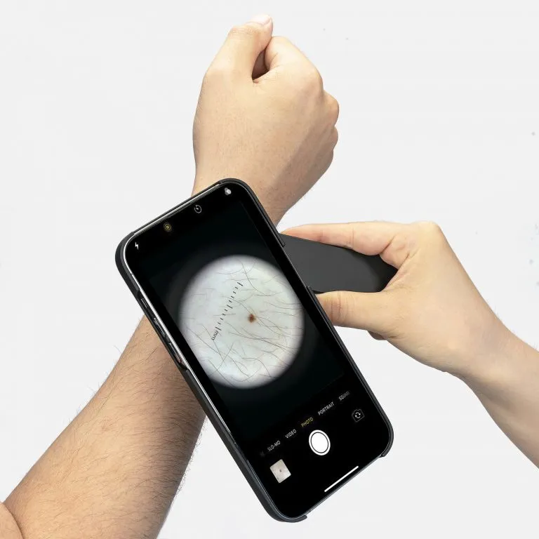
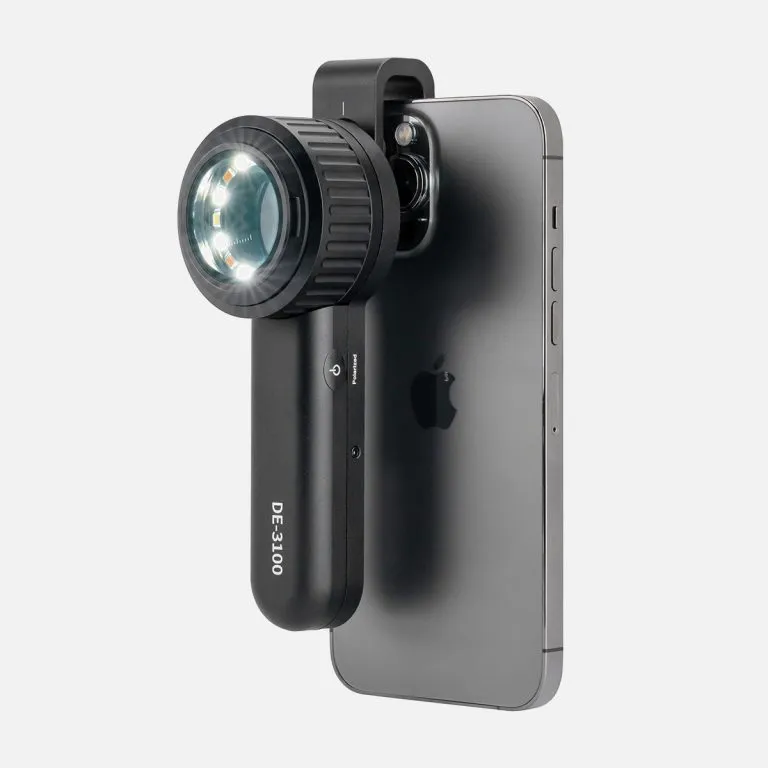
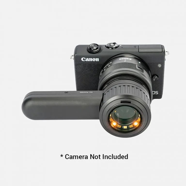
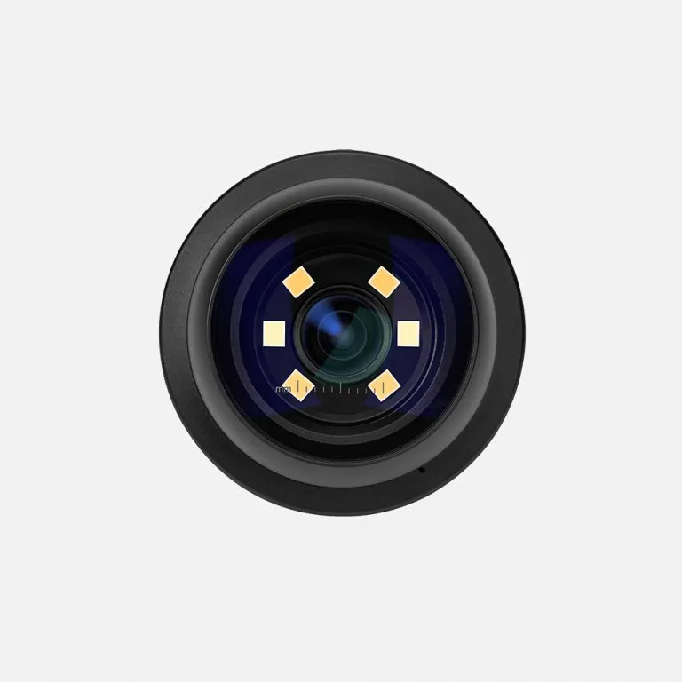
Our digital dermatoscope models (also known in international markets as dermatoscopio digital) leverage proprietary dermoscopic camera technology to capture distortion-free skin images. Unlike a traditional handheld lens, an electronic dermatoscope allows for real-time visualization on screens, making it the ideal dermatoscope camera for patient education and remote consultation.
Whether you need a high-end dermatoscope camera or a robust portable unit, IBOOLO ensures every device delivers clinical precision that meets the highest medical standards.
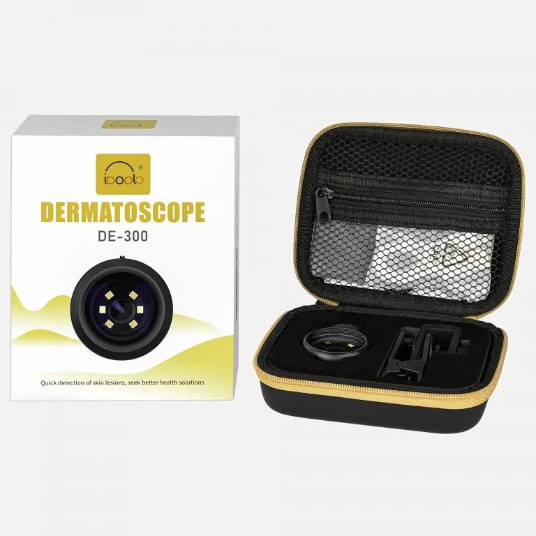
Many practitioners search for a dermatoscopio dermlite due to their industry reputation. IBOOLO offers a high-performance alternative that matches the optical clarity of leading brands but at a more accessible dermascope price. We focus on providing cheap dermatoscope options that do not compromise on medical quality—giving you the best dermatoscope price-to-performance ratio in the medical imaging market.
When you seek dermatoscopes for sale, reliability and after-sales support are as important as the dermatoscope cost. IBOOLO supports clinical practices worldwide with:
Explore our full collection of dermatoscopes for sale and get a quote today.
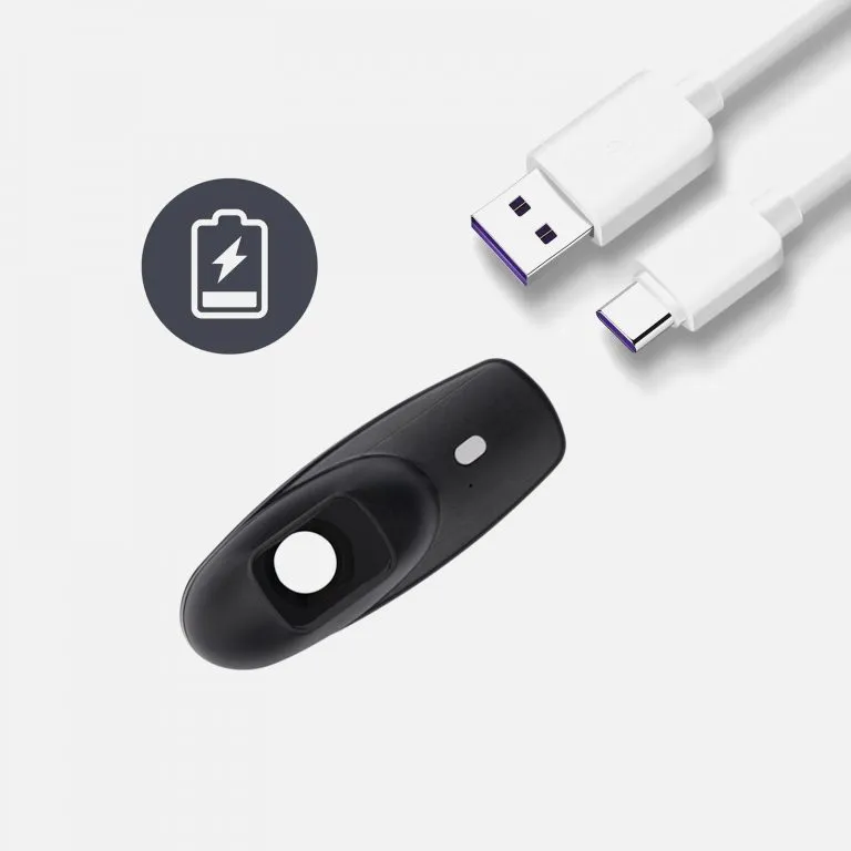
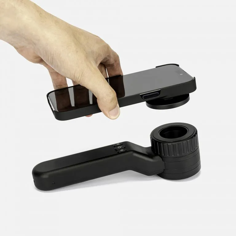
Get My Dermatoscope PriceOur China products supply hub couples world-class portability with elite precision, using seasoned expertise to develop high quality dermoscopy meaning for flawless skin visualization anywhere through compact size.
Our China products supply creates clinical quality Professional skin cancer dermoscopys enabling powerful skin magnification from anywhere through thoughtful craftsmanship.
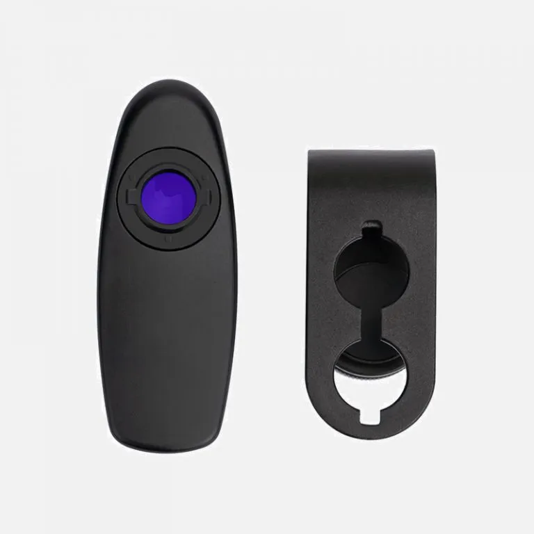
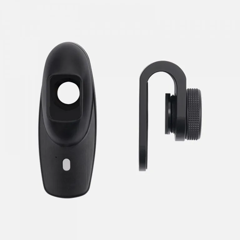
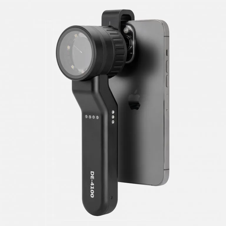
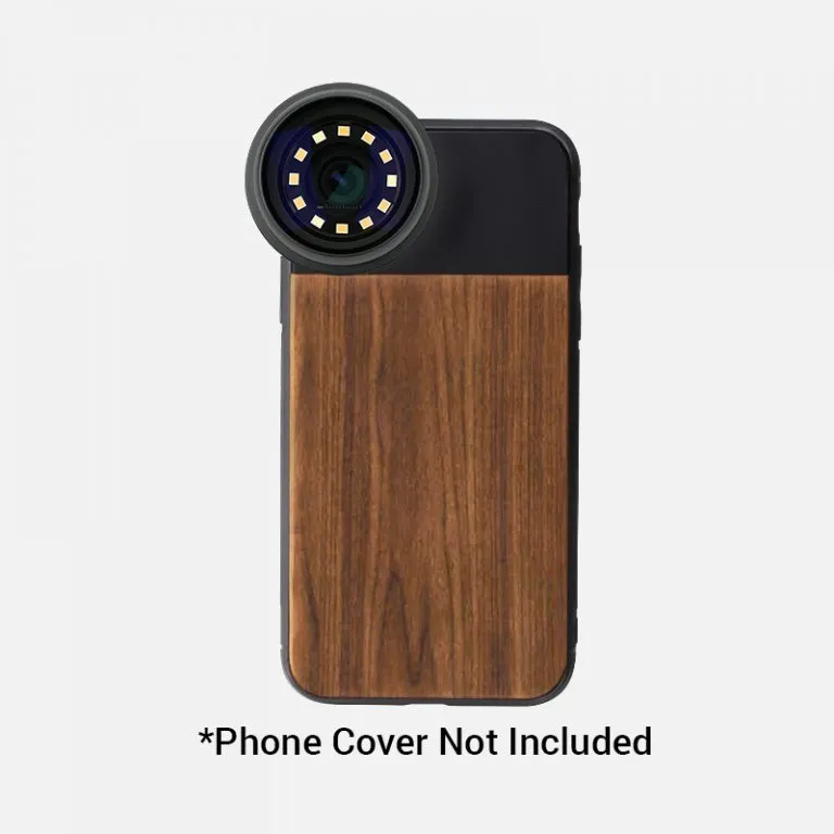
As an expert China products supply, we use exacting wholesale production methods to manufacture high-quality dermatoscope phone attachment solutions tailored for every customer.


 DE-500 Dermatoscope
1 × $399.00
DE-500 Dermatoscope
1 × $399.00 Subtotal: $399.00

We use cookies on this website to provide a better user experience. By continuing to browse the website, you are giving your consent to receive cookies on this site. For more details please read our Privacy Policy.