


Article
Dermascope Skin Analysis
Through dermoscopy, physicians are able to visualize slight alterations in the skin, such as blood vessels and pigmentation. It is first used for the screening of skin tumours, for the diagnosis of skin infections and inflammatory diseases, and for the monitoring of vascular skin lesions. This article provides a brief outlook on the application and…
Through dermoscopy, physicians are able to visualize slight alterations in the skin, such as blood vessels and pigmentation. It is first used for the screening of skin tumours, for the diagnosis of skin infections and inflammatory diseases, and for the monitoring of vascular skin lesions. This article provides a brief outlook on the application and effectiveness of dermoscopy. This article is helpful for readers to the know basic principles of how to use dermatoscopes correctly.
Introduction to Dermoscopy Examination
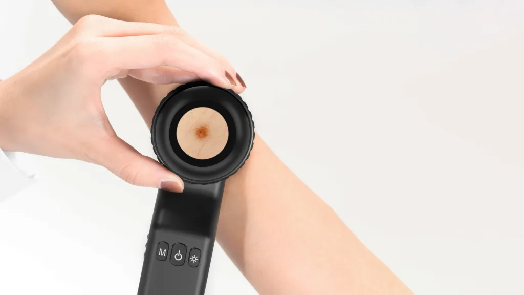
Dermoscopy is the use of a dermascope by a doctor to examine the skin of a patient. It needs only the full exposure of the examination area and no special preoperative preparation. For example, IBOOLO Dermatoscope is used to distinguish between benign pigmented nevi and malignant melanomas and to check for abnormal growth or malignancy.
Preparation for Dermoscopy
The patient will need to clean the skin prior to the examination, removing oils, dirt, make-up, hand-held patients. etc., dermatoscope, The so the examination that dermatoscope room the probe needs physician is to can first be clearly sterilised well visualise to lit the avoid so skin cross-infection that condition. between the Before doctor using can the clearly observe the skin condition. At the same time, the doctor needs to adjust the light and focus according to the observation needs.
Dermoscopy-related Equipment and Tools
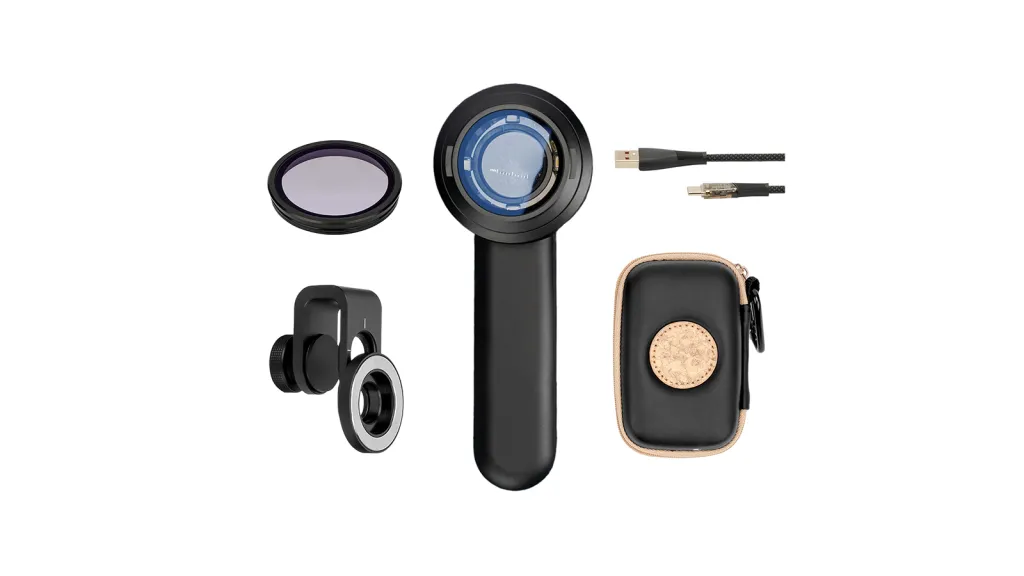
Dermascopes can be broadly categorised into digital dermoscopes and handheld dermoscopes. Handheld dermatoscopes are the most traditional and common type of dermatoscopy equipment. The DE-3100 and DE-4100 are part of IBOOLO’s professional handheld dermatoscopy series, with a magnification of 10X and four built-in different illumination modes.
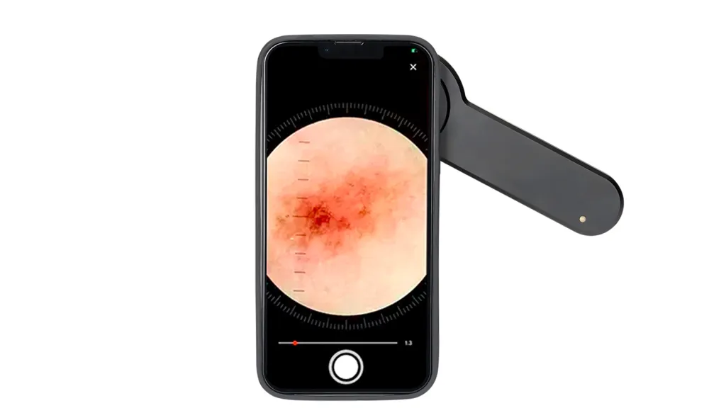
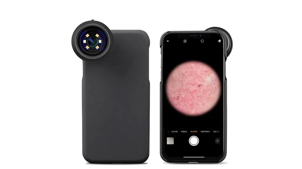
IBOOLO dermascope devices are now designed to be used with smartphone accessories. Examples include cases with threads as well as universal phone clips and magnetic rings. Users can use them with the accessories and save dermascope images through their mobile phones.
Steps in Dermoscopy
Prior to the dermoscopic examination, the patient will keep the area of skin under observation clean. The doctor should point the dermatoscope’s probe at the area to be examined, select the appropriate illumination mode and adjust the focus until the image is clear enough for observation.IBOOLO handheld dermatoscopes are packaged by default with a mobile phone clip and a magnetic ring, which allows the user to connect the dermatoscope to a mobile phone for image saving.
Analysis of Dermoscopic Images
Colour is one of the most visual and important features of a dermoscopic image, with different colours reflecting the depth, nature and composition of the skin lesion. Brown and black colours are usually associated with the distribution of melanin within the epidermis. Blue or grey, on the other hand, suggests that melanin is located in the dermis and is a potential marker of malignant melanoma.
Patterns are structural features in dermoscopic images that reflect the histological properties of a skin lesion, such as reticular patterns. Brown or black reticular structures are commonly found in benign nevi. Irregularities, fractures or thickening of the reticulation may indicate malignant lesions.
Structural analysis focuses on the borders, shape, and internal structure of the skin lesion. Benign lesions have clear, smooth borders. Malignant lesions often present fuzzy, irregular borders. Uniform structure is usually a sign of a benign lesion. Uneven, disorganised structures suggest malignant potential.
Dermoscopic image analysis can assist the physician in the early identification of malignant lesions such as melanoma, basal cell carcinoma, and squamous cell carcinoma by providing a detailed view of the colours, patterns, and structures. Dermoscopy improves diagnostic sensitivity and specificity compared to visual observation.
Clinical Significance of Dermatological Microscopy
Through dermoscopy, doctors can distinguish between harmless and dangerous skin growths and enhance the diagnostic precision, rate which for may early decrease stage the melanoma chances can of be misdiagnosis 90% or or underdiagnosis. better, The while five the year survival survival rate for advanced melanoma is significantly worse. Other skin cancers are also highly curable if treated surgically at an early stage with a cure rate of nearly 100%.
Limitations and Challenges of Dermoscopy
This is because dermoscopy enhances the early diagnosis precision through magnification of skin features and visualization of the skin surface and its superficial structures in comparison to the conventional visual assessments. Compared to other techniques, imaging such as skin ultrasound and optical coherence tomography, it has the advantages of being portable, relatively to cheap, display and information easy from to deeper use. tissues, However, and it having has specific the and disadvantages sensitive of signals being that operator are dependent, determined unable by optical methods. These issues can be solved by increasing the level of training of physicians, incorporating other diagnostic methods (biopsy and ultrasound), and enhancing the equipment technology, which may include higher resolution and multimodal imaging.
Future Developments in Dermoscopy
Future developments in dermoscopy are focused on technological progress and skill development. AI can enhance the ability of doctors to analyze dermoscopy images quickly and accurately, detect malignant lesions and decrease the probability of misdiagnosis. However, telemedicine and digital technologies are also expanding the front door of the doctor’s office to the internet, enabling image sharing, specialist cooperation, and tele-diagnosis, especially in areas with limited access to healthcare providers. To fully take advantage of these technologies, doctors will need to resources continue and to gaining develop hands-on their experience. skills, The for use instance of by technology attending and training specialisation sessions, will using increase the the internet role and of other electronic dermoscopy in diagnosis, prevention and management of health, thus making precision medicine available to more people.
Dermoscopy and Skin Health Management
Dermoscopy is a non-invasive, rapid and highly effective tool for the evaluation of skin lesions. It can help physicians differentiate between benign and malignant skin lesions and is especially important in the early detection of malignant tumours such as melanoma. In skin health monitoring, dermoscopy not only helps doctors to make an accurate diagnosis, but also provides a basis for ongoing follow-up and evaluation.
Dermascope Skin Analysis: Precision Mapping and Clinical Interpretation
In the realm of modern dermatology, dermascope skin analysis has transitioned from a specialized clinical procedure to an essential standard for skin health monitoring. By utilizing advanced optical systems to bypass the refractive index of the stratum corneum, this analytical technique allows for the visualization of subcutaneous structures that remain invisible during standard visual inspections. Whether for professional oncological screening or proactive personal monitoring, understanding the nuances of dermascopic imaging is key to early intervention.
The Core Principles of Dermascope Skin Analysis
The effectiveness of skin analysis using a dermatoscope is rooted in high-definition magnification combined with controlled illumination. Unlike a standard magnifying glass, a professional dermatoscope like the IBOOLO DE-4100 utilizes cross-polarization to eliminate surface glare, providing a clear window into the dermal-epidermal junction. This allows clinicians to perform a detailed analysis of pigmentation distribution and vascular morphology without the interference of ambient light reflections.
Key Diagnostic Parameters in Skin Analysis
When performing a dermascope skin analysis, experts evaluate three primary visual cues to determine the nature of a lesion. These observations are critical for differentiating between benign nevi and malignant growths.
1. Chromatic Evaluation (Color Patterns)
The color observed during an analysis provides immediate clues regarding the depth of the melanin. In a standard analysis: - Black and dark brown tones typically indicate melanin in the upper epidermis. - Slate-gray or blue hues suggest deeper dermal involvement, which is a significant marker in the analysis of potentially malignant melanoma.
2. Architectural Symmetry and Patterns
A structured dermascope skin analysis focuses heavily on the regularity of patterns. Benign lesions usually exhibit a symmetric reticular (mesh-like) pattern. In contrast, an analysis that reveals a "starburst" pattern or a multi-component pattern—where different structures coexist in one lesion—often warrants a closer clinical investigation or a biopsy.
3. Border and Structural Integrity
Analyzing the periphery of a lesion is essential. Smooth, well-demarcated borders are characteristic of benign structures. During a professional analysis, any observation of irregular, "moth-eaten" edges or peripheral streaks (pseudopods) is documented as a high-risk indicator for skin cancer progression.
Optimizing the Workflow with IBOOLO Technology
To achieve a reliable dermascope skin analysis, the hardware must facilitate a seamless workflow. IBOOLO devices are engineered to support this process through several technical advantages:
- High-Resolution Optics: 10x magnification with premium Japanese glass ensures edge-to-edge clarity during every analysis session.
- Dual Illumination Modes: Clinicians can toggle between polarized light for deep vascular analysis and non-polarized light for superficial texture evaluation.
- Smartphone Linkage: By utilizing universal adapters, IBOOLO enables teledermatology, allowing analysis images to be captured in 4K and shared instantly for remote consultation.
Step-by-Step Guide to Performing an Accurate Analysis
- Skin Preparation: Ensure the target area is free of oils and cosmetics that might obscure the micro-structures.
- Mode Selection: For initial screening, use non-polarized light to assess surface scales. Switch to polarized mode for a deeper analysis of the pigment network.
- Focus Adjustment: Utilize the fine-focus ring of the IBOOLO device to achieve a sharp image of the dermal layer.
- Documentation: Capture high-quality images for longitudinal analysis, comparing changes in size, color, or pattern over time.
Frequently Asked Questions
How does dermascope skin analysis improve diagnostic accuracy?
Research indicates that integrating dermoscopy into skin examinations can improve the sensitivity of melanoma detection by over 35% compared to visual observation alone.
Can I perform a dermascope skin analysis at home?
While IBOOLO pocket dermatoscopes are user-friendly enough for personal monitoring, a definitive analysis and diagnosis should always be conducted by a qualified healthcare professional.
What is the role of AI in future skin analysis?
The integration of AI algorithms with digital dermascope skin analysis is the next frontier, providing clinicians with automated pattern recognition to assist in identifying suspicious lesions faster.
Recommended reading
Wholesale Dermascope Vs Dermatoscopes Products Supply by Company in China - IBOOLO
We are a leading China-based dermascope vs dermatoscope products supply capable of providing wholesale lamps designed around our clients' specific needs. Our flexible products supply process ensures tailored solutions.
What’s New in Dermatoscopes 2024 – IBOOLO
We are constantly improving our dermascopes and trying to offer innovative & affordable dermascopes. We want to leverage our expertise in optical fields, to create medical lenses that benefit humanity.
Dermascope vs Dermatoscope: Surpassing the Magnifying Glass - IBOOLO
IBOOLO: Dermascopes vs. Dermatoscopes - Surpassing the Limitations of the Magnifying Glass to Enable Precise Skin Evaluations and Enhanced Patient Outcomes


