


Article
Dermoscopy of Seborrheic Keratosis Dermatitis
Seborrheic keratosis dermatitis is a common skin hyperplasia. It is often mistaken for a disease such as skin cancer because of its appearance that looks like warts, precancerous skin growths, or skin cancer. Dermoscopy of seborrheic keratosis dermatitis is crucial to identify seborrhei keratosis from other types of skin diseases. What is Seborrheic Keratosis Dermatitis?Seborrheic…
Clinical Excellence: Advanced Dermoscopy of Seborrheic Keratosis in Early and Irritated Stages
In modern dermatology, the dermoscopy of seborrheic keratosis (SK) is far more than a routine check; it is a critical safeguard against misdiagnosing malignant tumors. Seborrheic keratosis, while inherently benign, exhibits a morphological plasticity that can mimic melanoma or basal cell carcinoma (BCC), especially when presented in its early developmental phase or under inflammatory irritation.
As a global leader in optical diagnostic tools, IBOOLO provides this deep-dive clinical analysis to empower clinicians in identifying subtle micro-structures and complex vascular patterns that define the evolution of seborrheic keratosis.
1. The Diagnostic Threshold of Early Seborrheic Keratosis Dermoscopy
The primary challenge in early seborrheic keratosis dermoscopy lies in the absence of mature "pasted-on" plaques. In these incipient lesions, the hallmark milia-like cysts may be microscopic or entirely absent.
To achieve a precise diagnosis, clinicians must focus on "Nascent Patterns":
- Faint Fingerprint-like Structures: These represent early acanthosis and are often the first sign of epidermal thickening before the lesion becomes verrucous.
- Moth-eaten Borders: This specific concave indentation of the lesion margin is a highly predictive sign in early seborrheic keratosis dermoscopy, distinguishing it from the sharp, expansive borders of a melanocytic nevus.
- Milia-like Cysts (Polarized View): Under polarized light, even in early stages, tiny crystalline white dots (keratin pearls) can often be detected deep within the invaginations.
2. Navigating the Inflammatory Shift: Irritated Seborrheic Keratosis Dermoscopy
When a lesion undergoes trauma or friction, the resulting irritated seborrheic keratosis dermoscopy profile shifts significantly. Inflammation can induce a "pseudosarcomatous" appearance, masking classic structural clues.
Strategic indicators for irritated lesions include:
- The "Red Halo" Phenomenon: Diffuse erythema surrounding the base of the lesion, often accompanied by superficial scales or "Squamous Eddies" (concentric circles of keratin).
- Masking of Cysts: In irritated seborrheic keratosis dermoscopy, milia-like cysts may be obscured by inflammatory exudate. Clinicians should inspect the periphery for "remnant structures" to confirm the benign origin.
- Structural Integrity: Despite the irritation, the underlying "cerebriform" (brain-like) pattern usually remains intact at the core, providing a reliable diagnostic anchor.
3. Vascular Morphology: Decoding Seborrheic Keratosis Dermoscopy Vessels
Vascular analysis is the ultimate differentiator. In inflamed or non-pigmented lesions, seborrheic keratosis dermoscopy vessels provide the final clue to excluding BCC or melanoma.
Key vascular archetypes to identify:
- Hairpin Vessels with White Halos: This is the most specific pattern for SK. These U-shaped looped capillaries are surrounded by a distinct whitish perivascular halo, representing the keratinized tissue.
- Regularity vs. Chaos: Unlike the disorganized, polymorphous vessels of melanoma, seborrheic keratosis dermoscopy vessels are distributed with remarkable symmetry and monomorphism across the lesion surface.
- Dotted Vessels: In some variants, regularly arranged red dots may dominate. However, they lack the "cluster" arrangement typical of squamous cell carcinoma.
Differential Diagnosis: SK vs. Malignant Mimickers
A professional dermatoscope like the IBOOLO DE-4100 allows for the clear visualization of these differences. The table below outlines the critical thresholds:
| Feature | Seborrheic Keratosis (SK) | Malignant Melanoma |
|---|---|---|
| Symmetry | High (Organized Patterns) | Low (Structural Chaos) |
| Vascular Type | Hairpin / Looped | Polymorphous / Corkscrew |
| Specific Sign | Comedo-like openings | Irregular Pigment Network |
Precision Diagnostics: The IBOOLO Advantage in SK Screening
Visualizing seborrheic keratosis dermoscopy vessels in an irritated state requires superior light control. IBOOLO’s dual-polarization technology is engineered to cancel out surface glare from thick keratin, revealing the underlying vascular architecture that non-polarized lenses often miss.
By utilizing 4K high-resolution imaging, our devices allow clinicians to perform early seborrheic keratosis dermoscopy with unprecedented clarity, capturing the minute fingerprint-like structures that lead to confident, non-invasive diagnoses and reduced biopsy rates.
Recommended reading
Affordable Polarized Light Dermoscopy Selection from Top China Products Supply - IBOOLO
As a leading affordable polarized light dermoscopy products supply in China, over 11+ years experience allows us to packed professional grade viewing into easy-to-carry mobile devices.
Dermoscopy of Malenoma – IBOOLO
What is malignant melanoma?Malignant melanoma is a serious skin cancer that starts in melanocytes. It is also known as cutaneous melanoma. This skin cancer is much dangerous due to its rapidly spread to other organs if it is not controlled at an early stage.Melanoma can appear in anywhere on the skin, but for people with...
Dermoscopy of Dermatofibroma – IBOOLO
A dermatofibroma is a common benign bump in the skin.Dermatofibroma is usually harmless and typically appears on the lower legs. Dermatofibroma is easily mistaken from other skin tumors due to its appearance. This brings much difficult to diagnose. Hence, how to exactly diagnose dermatofibroma is special important to dermatology filed. What is dermatofibroma...
Seborrheic keratosis dermatitis is a common skin hyperplasia. It is often mistaken for a disease such as skin cancer because of its appearance that looks like warts, precancerous skin growths, or skin cancer. Dermoscopy of seborrheic keratosis dermatitis is crucial to identify seborrhei keratosis from other types of skin diseases.
What is Seborrheic Keratosis Dermatitis?
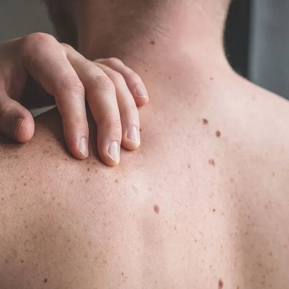
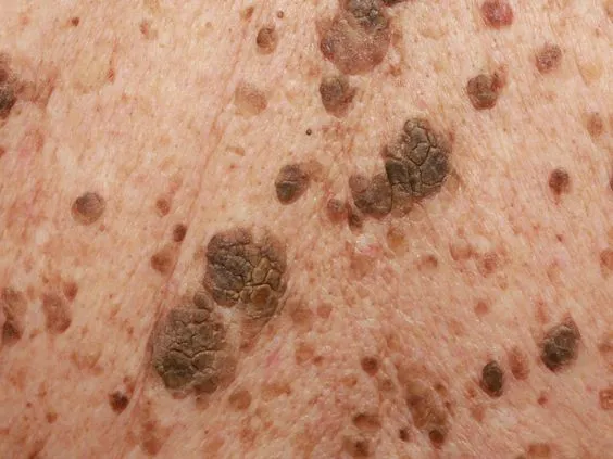
Seborrheic keratosis dermatitis is a kind of benign epidermal hyperplasia caused by keratinocyte hyperplasia. Seborrheic keratosis dermatitis is a type of non-cancerous benign of skin disease. Seborrheic keratosis dermatitis is harmless.
Seborrheic keratosis dermatitis is known as senile warts, senile spots, also known as basal cell papilloma. Because it mainly occurs in adults over the age of 40, it often appears as people grow older.
What are the Clinical Feature of Seborrheic Keratosis Dermatitis?
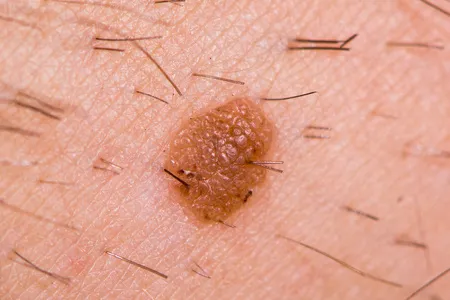
Seborrheic keratosis dermatitis is painless and it usually appears brown, black, or light tan. Its growth appears waxy or scaly and is slightly raised. They can gradually appear on various parts of the body, mostly on the face, neck, chest, or back.
Why is It Necessary to Use a Dermoscopy of Seborrheic Keratosis Dermatitis?
Dermatoscope is a non-invasive technique that allows dermatologists to closely examine seborrheic keratosis dermatitis more accurately and precisely. Especially dermoscopy of seborrheic keratosis dermatitis greatly enhance the vision of some locations that hard-to-reach by naked eyes, such as details in lesions. Dermatoscope magnifys and brightens shapes and structures of lesions. Dermoscopy of seborrheic keratosis dermatitis increases the confidence of doctors and patients about the skin disease and avoids unnecessary anxiety and treatment. So it is really necessary to use a dermoscopy of seborrheic keratosis.
Typical Features of Dermoscopy of Seborrheic Keratosis Dermatitis
dermoscopy plays a crucial role in identifying seborrheic keratosis dermatitis. There are some typical features of dermoscopy of seborrheic keratosis dermatitis as below:
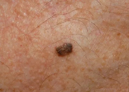
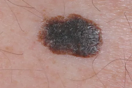
Special pattern: Typical “gyrigrain” or “fat-like” pattern.
Hair follicle openings: Visible hair follicle openings.
Structure: Edge ring structure, Light brown fingerprint-like parallel structures.
Prominent blood vessels: In some forms of seborrheic keratosis dermatitis, a halo of lobules surrounds tiny, hairpin-like capillaries.
Miliary cysts: These cysts may appear as small white stars or larger, yellowish turbidity.
Other features like: cracks/ridges, blue-gray balls, irregular crypts, weak or pseudo-network.
Dermoscopy of seborrheic keratosis dermatitis is very helpful and reliable for distinguish seborrheic keratosis dermatitis from other skin diseases.
How to differentiate between seborrheic keratosis dermatitis and melanoma?
Seborrheic kearatosis dermatitis will not transfer into melanoma. But both of seborrheic keratosis dermatitis and melanoma can be brown or black color, so the two can be easily be mistake from each other.
There are some important differences between seborrheic keratosis dermatitis and melanoma, from their numbers, appearances, locations causes, etc.
Comparison the apearances of seborrheic keratosis dermatitis and melanoma
Numbers: Seborrheic keratoses dermatitis: Seborrheic keratoses dermatitis often appear in numbers of two or more
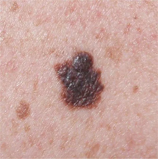
Melanoma: Melanoma is usually appear in single.
Shapes: Seborrheic keratoses dermatitis: Seborrheic keratoses dermatitis usually shows round or oval shaped.
Melanoma: Irregular shape, asymmetry in shape is the typical features of melanoma.
Colors: Seborrheic keratoses dermatitis: Seborrheic keratoses dermatitis colors in light tan, brown, or black.
Melanoma: Melanoma is commonly display multiple colors like pink, red, white, blue or mixed color within the same one.
Size: Seborrheic keratosis dermatitis: Seborrheic keratosis dermatitis varies in size from very small to big, and its size will not changed as time goes on.
Melanoma: Melanoma is in bigger size than 1/4 inch, and its size will grow over time.
Surface: Seborrheic keratosis dermatitis: Seborrheic keratosis dermatitis has waxy or scaly surface, slightly elevated above the skin surface.
Melanoma: Melanoma tends to be smooth with blurred, ragged border.
Pain: Seborrheic keratosis dermatitis:Seborrheic keratosis dermatitis is painless
Melanoma: Some of melanoma feel hurt, while some of melanoma feel no any pain or discomfort.
Evolving: Seborrheic keratosis dermatitis:Seborrheic keratosis dermatitis maintains the same always.
Melanoma: Melanoma may looks different from its beginning, and it may change in its shape, size or color.
Comparison the locations of seborrheic keratosis dermatitis and melanoma
Seborrheic keratosis dermatitis: Seborrheic keratosis dermatitis mostly displays on the face, neck, chest, or back.
Melanoma: Melanoma Melanoma can appear in anywhere on the skin,mostly on chest, black, legs, arms, face, necks, and even eyes.
Comparison the causes of seborrheic keratosis dermatitis and melanoma
Causes: The primary risk factor for seborrheic keratoses is age. Other risk factors include: sunburn, skin irritation and friction, pregnancy, hormone therapy, some medications, genetic mutation, a family history of seborrheic keratosis
How is seborrheic keratosis dermatitis diagnosed?
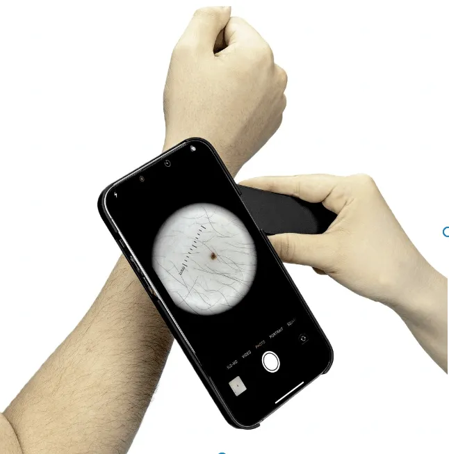
To diagnose seborrheic keratosis dermatitis, skin doctors will get information from your family history of skin disease and take observation of it through a vision aiding tool called dermatoscope. Dermatoscope is a small handheld lighted medical microscope that allows a more precise and deeper view of skin diseases by high magnification and a powerful glare-free lighting system. If it is necessary, a biopsy will be needed for seborrheic keratosis.
People usually also take dermoscopy of seborrheic keratosis dermatitis for self-examination on skin. Any unusual findings or changes occur, have dermatologist checked for a further evaluation. Dermoscopy of seborrheic keratosis dermatitis plays a significant role in physical exam.
Application of dermoscopy of seborrheic keratosis dermatitis
A dermoscope is a main device used to examine skin diseases, like seborrheic keratosis dermatitis. In the diagnosis and treatment of seborrheic keratosis dermatitis, dermoscopy of seborrheic keratosis dermatitis is widely used in the following aspects:
Monitoring: For patients who have already been diagnosed with seborrheic keratosis dermatitis, dermoscopy can be used to monitor the whole process as time goes on. If any changes happen, a further step should be taken.
Feedback: Skin doctor can compare images from dermoscopy of seborrheic keratosis dermatitis over different times to assess the effectiveness of the treatment of seborrheic keratosis dermatitis and then decide if the treatment needs to be adjusted or not.
Treatment aid: When treating seborrheic keratosis dermatitis, images can be clearly and precisely showed by dermoscopy of seborrheic keratosis dermatitis. It greatly enhanced the patience of skin doctors and patients.
Seborrheic keratosis dermatitis is a harmless skin disease which will not cause skin cancer. But skin doctors should have it accurately diagnosed by dermoscope. Dermoscopy of seborrheic keratosis dermatitis is very important to differentiate seborrheic keratosis dermatitis from other skin diseases. Hence, skin doctors can remove seborrheic keratoss surely and safely for some aesthetic reasons.
It is vital to develop the habit of use of dermoscopy of seborrheic keratosis dermatitis. In addition, paying more attention to a regular skin examination is also necessary in our daily life. People should remain vigilant at all time to keep the health of the skin.


