

-
×
 DE-3100 Dermatoscope
1 × $499.00
DE-3100 Dermatoscope
1 × $499.00
Subtotal: $499.00
Message reply within 1 day
Free national shipping
100% satisfaction
2 years warranty
IBOOLO DE-3100 is a clinical-grade polarized dermatoscope featuring hybrid toggle modes. Master polarized dermoscopy for early melanoma detection and skin imaging.
The IBOOLO DE-3100 represents a new standard in clinical skin imaging. As a high-performance polarized dermatoscope, it is engineered specifically to meet the rigorous demands of modern dermatology. By integrating advanced polarized dermoscopy technology, the DE-3100 allows practitioners to see deeper, clearer, and more accurately, ensuring that no subtle lesion goes unnoticed.
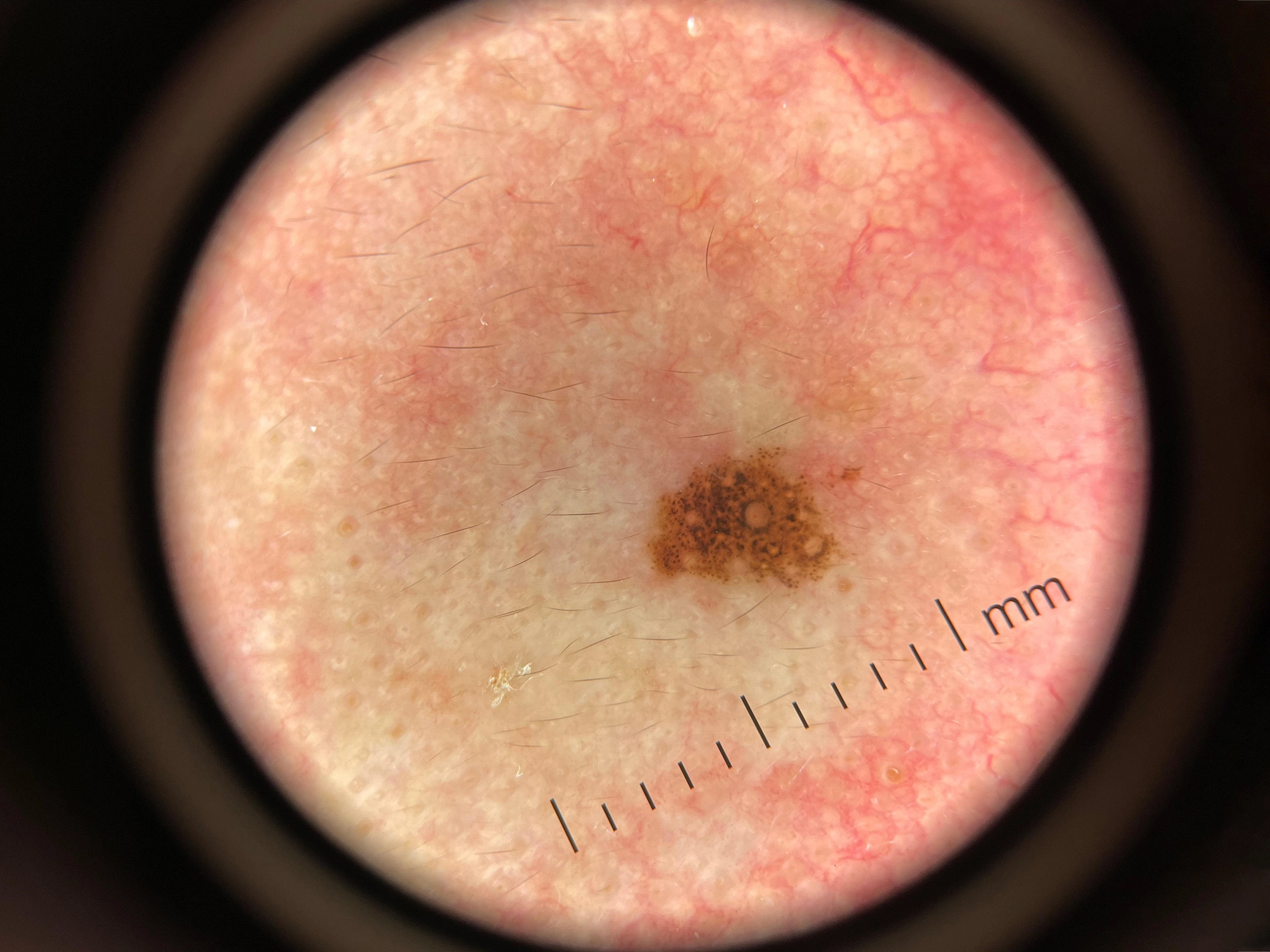
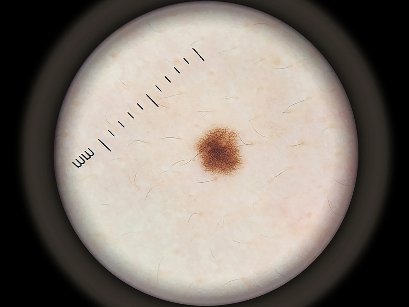
Traditional non-polarized dermatoscopy requires a liquid interface (immersion oil) and direct skin contact to eliminate surface reflection. However, the DE-3100 polarized dermatoscope utilizes cross-polarization filters to block backscattered light from the stratum corneum. This allows for polarized light dermoscopy to be performed without skin contact, providing a crystal-clear window into the deeper layers of the dermis.
By using the DE-3100, clinicians can visualize vascular patterns, crystalline structures, and collagen changes that are often invisible under standard light. This makes polarized dermoscopy an essential technique for the early diagnosis of both melanoma and non-melanoma skin cancers.
The DE-3100 is not just a tool; it is a comprehensive diagnostic solution. Its design reflects a decade of optical expertise at IBOOLO, focusing on three core pillars: Clarity, Versatility, and Reliability.
One of the most powerful features of the DE-3100 is its ability to toggle between polarized and non-polarized modes at the touch of a button. While polarized dermoscopy excels at showing deep vascular structures, non-polarized mode is still valuable for seeing superficial features like comedo-like openings or milia-like cysts. The DE-3100 gives you the best of both worlds in one device.
Equipped with a high-definition achromatic lens system, the DE-3100 dermatoscope provides 10x magnification with zero edge-to-edge distortion. This ensures that every polarized light dermoscopy session delivers high-fidelity images essential for dermatological documentation and telemedicine.
Designed for the "physician on the move," the DE-3100 is lightweight yet durable. Its ergonomic grip reduces hand fatigue during long clinical sessions, while its universal smartphone compatibility allows it to be used as a mobile polarized dermoscopic camera.
Choosing the right polarized dermatoscope requires understanding how light interacts with skin anatomy. Below is a technical breakdown of how the DE-3100 enhances your diagnostic capabilities.
| Optical Characteristic | Polarized Mode (DE-3100) | Traditional Non-Polarized |
|---|---|---|
| Reflection Control | Cross-polarization (Zero Glare) | Requires Immersion Fluid |
| Visual Depth | Deep Dermal Structures | Superficial Epidermal Focus |
| Vascular Visualization | Highly Detailed (Red/Pink patterns) | Often Obscured by Glare |
| Crystalline Structures | Visible (Pathognomonic for BCC/Melanoma) | Invisible |
| Clinical Workflow | Fast, Non-contact, Hygienic | Slower, Contact-required |
The versatility of polarized dermoscopy extends beyond simple mole checks. The DE-3100 is a multi-disciplinary tool used in various medical fields:
When you invest in an IBOOLO polarized dermatoscope, you are investing in a medical device that meets global safety and quality standards. IBOOLO is an ISO 13485:2016 certified manufacturer. The DE-3100 is FDA registered and CE marked, ensuring that it is suitable for professional use in hospitals, clinics, and research institutions worldwide.
Q: What is the main advantage of a polarized dermatoscope?
A: A polarized dermatoscope allows for non-contact examination and reveals deeper skin structures (like blood vessels and collagen) that are hidden by surface glare under traditional light.
Q: Does the DE-3100 require immersion oil?
A: No. Thanks to advanced polarized dermoscopy technology, the DE-3100 eliminates reflections electronically through its lens system, making immersion oil unnecessary for most clinical exams.
Q: Can I use the DE-3100 with my smartphone?
A: Yes. The DE-3100 is designed with universal compatibility, allowing it to clip onto any smartphone to become a high-resolution digital polarized dermatoscope.
Q: Why is polarized light better for melanoma detection?
A: Polarized light dermoscopy makes "crystalline structures" (white shiny lines) visible. These structures are highly specific indicators of invasive melanoma and basal cell carcinoma.
Our China products supply hub couples world-class portability with elite precision, using seasoned expertise to develop high quality dermoscopy meaning for flawless skin visualization anywhere through compact size.
Our China products supply creates clinical quality Professional skin cancer dermoscopys enabling powerful skin magnification from anywhere through thoughtful craftsmanship.
As an expert China products supply, we use exacting wholesale production methods to manufacture high-quality dermatoscope phone attachment solutions tailored for every customer.
High Quality Rechargeable LED DE-3100 Dermatoscope


$499.00
| Material | Aluminum & Glass |
| Optical Design | All glass, 4 elements 3 groups, anti-reflection coating |
| Lens Diameter | 32mm(front); 25mm(rear) |
| Magnification | 10x |
| Distortion | 8% |
| Polarization | Cross Polarization |
| Resolution | 300 LP/MM (Axis) 280 LP/MM (Edges) |
| LED | 30 LEDs, 18 polarization, 6 non polarization, 6 amber polarization |
| Battery Capacity | 1000mAh Lithium ion |
| Charging | USB-C |
| Working Time | 6-8 hours |
| Focus Distance | 12-20mm |
| Dimension | Φ53mm*H35mm*L136.5mm |
| Weight | 187g |
$499.00


$3.90 – $14.90

$15.90
$3.90 – $14.90
$15.90

It’s a handheld dermatoscope, but it contains a universal phone adapter meaning convenient to connect with any smartphone or tablet to capture images. Build with 4K resolution optics,polarized light, 10X magnification and 25mm wide field of view — the DE-3100 is perhaps the most super value dermatoscope we’ve ever seen. The visual filed is smaller(22%) than DE-4100, but the weight is 40% lighter.
Made from premium multi-coated and multi-element optics, the imaging performance is leading in the market. Optimized for the latest iPhone, rank only next to DE-4100.
Naked eye detection. 25mm visual field of view.

In polarization mode, the polarization filter becomes activated and absorbs the surface light reflection (no immersion fluid required). It allows you to examine the colours, shapes and texture features of the skin lesion more clearly, more precise and more detailed.

Built with polarized & non-polarized light, the DE-3100 offers two levels of color spectrum control to enhance imaging of deeper pigmentation.

The magnet attachment gives the ability to attach on smartphone and take pictures in 5 seconds.
Best part? It compatible with all phone in the market, with a universal lens clip or a phone cover for easy on and off.
Focus adjustable. It’s able to adjust working distance of different phone cameras. This enables the best image capture effect to be achieved.


There’s an influx of dermatoscope on the market. Our devices are remarkable blend of pro-level features and affordable price. Their premium optics and efficient LED system delivers sharp & precise images.


| Product | LENS DIAMETER | OPTICAL DESIGN | MAGNIFICATION | LIGHTING SYSTERM | BATTERY | CONNECTABILITY | ADJUSTABLE | COST |
|---|---|---|---|---|---|---|---|---|
DE-4100 |
48mm/32mm | 4 elements 3 groups | 10 times | 22 LEDs, polarise, non polarise, amber light | 1000mAh | naked eye, phone, tablet, digital camera | focus & brightness | $699.00 |
DE-3100 |
32mm/25mm | 4 elements 3 groups | 10 times | 30 LEDs, polarise, non polarise, amber light | 1000mAh | naked eye, phone, tablet, digital camera | focus | $499.00 |
DE-500 |
25mm/17mm | 4 elements 3 groups | 10 times | 16 LEDs, polarise, non polarise, UV light | 200mAh | smartphone & tablet | brightness | $399.00 |
DE-400 |
46mm/17mm | 4 elements 3 groups | 10 times | 12 LEDs, polarization, non polarization | 200mAh | smartphone & tablet | non adjustable | $179.00 |


Only logged in customers who have purchased this product may leave a review.

Melanoma is one of the most dangerous types of skin cancer. Although it is less common than other skin cancers, it causes the majority of skin-cancer related deaths. The main…

Optical clarity is the cornerstone of accurate dermatological assessment. The achromatic lens system used in devices such as the IBOOLO DE-4100 Dermatoscope relies on precision multi-layer coatings to minimize chromatic…

The release of the iPhone 17 Pro Max marks a significant leap in mobile imaging technology. With its advanced sensor upgrades and computational photography, it offers unprecedented potential for clinical…
DE-3100 Dermatoscope
1 × $499.00 Subtotal: $499.00

We use cookies on this website to provide a better user experience. By continuing to browse the website, you are giving your consent to receive cookies on this site. For more details please read our Privacy Policy.


Alan Rosenbach Canada
–
Canada
–
This dermatoscope is excellent. The attachment to the phone is a home run. Viewing is much easier. Great company with great communication